. 2007 Sep;81(17):9307-18.
doi: 10.1128/JVI.00900-07. Epub 2007 Jun 20.

Relevance of the interaction between alphaherpesvirus UL3.5 and UL48 proteins for virion maturation and neuroinvasion

Affiliations

Relevance of the interaction between alphaherpesvirus UL3.5 and UL48 proteins for virion maturation and neuroinvasion

Walter Fuchs et al. J Virol. 2007 Sep.

Abstract

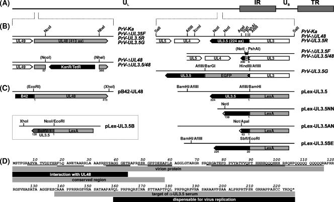
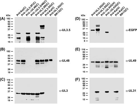
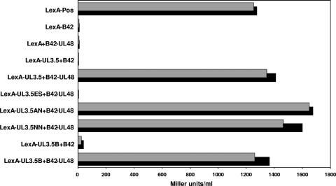
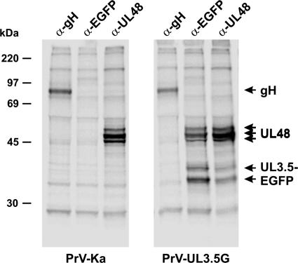
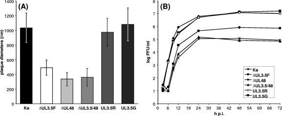
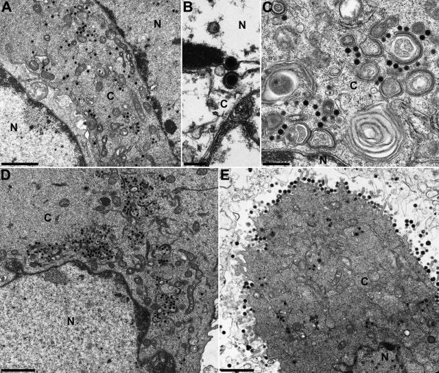
The UL3.5 and UL48 genes, which are conserved in most alphaherpesvirus genomes, are important for maturation of pseudorabies virus (PrV) particles in the cytoplasm of infected cells (W. Fuchs, B. G. Klupp, H. J. Rziha, and T. C. Mettenleiter, J. Virol. 70:3517-3527, 1996; W. Fuchs, H. Granzow, B. G. Klupp, M. Kopp and T. C. Mettenleiter, J. Virol. 76:6729-6742, 2002). In bovine herpesvirus 1 (BoHV-1), the homologous gene products pUL3.5 and pUL48 have been demonstrated to interact physically (N. Lam and G. Letchworth, J. Virol. 74:2876-2884, 2000). Moreover, BoHV-1 pUL3.5 partially complemented a pUL3.5 defect in PrV (W. Fuchs, H. Granzow, and T. C. Mettenleiter, J. Virol. 71:8886-8892, 1997). By using coimmunoprecipitation and yeast two-hybrid studies, we observed a similar interaction between pUL3.5 and pUL48 of PrV, as well as a heterologous interaction between the PrV and BoHV-1 gene products. The relevant domain could be confined to the first 43 amino acids of PrV pUL3.5. Unlike its BoHV-1 homologue, PrV pUL3.5 is processed by proteolytic cleavage, and only an abundant 14-kDa fragment consisting of amino acids 1 to >or=116 could be detected by peptide mass fingerprint analysis of purified wild-type PrV particles, which also contain the pUL48 tegument component. To determine the biological relevance of the protein-protein interaction, pUL3.5-, pUL48-, and double-negative PrV mutants were analyzed in parallel. All deletion mutants were replication competent but exhibited significantly reduced plaque sizes and virus titers in cultured rabbit kidney cells compared to wild-type and rescued viruses, which correlated with a delayed neuroinvasion in intranasally infected mice. Remarkably, the defects of the double-negative mutant were similar to those of pUL48-negative virus. Electron microscopy of cells infected with either deletion mutant revealed the retention of naked nucleocapsids in the cytoplasm and the absence of mature virus particles. In summary, our studies for the first time demonstrate the relevance of the pUL3.5-pUL48 interaction for secondary envelopment of an alphaherpesvirus, give a molecular basis for the observed trans-complementation between the PrV and BHV-1 pUL3.5 homologs, yield conclusive evidence for the incorporation of a proteolytically processed pUL3.5 into PrV virions, and demonstrate the importance of both proteins for neuroinvasion and neurovirulence of PrV.


Figures

FIG. 1.
FIG. 1.
Maps of virus mutants and plasmids. (A) The PrV genome, consisting of long (UL) and short (US) unique regions and inverted repeat sequences (IR, TR), is depicted (B) Enlarged sections show the viral ORFs (pointed rectangles) within the analyzed genome regions. Recombinant viruses were generated by mutagenesis in E. coli of an infectious full-length clone of the PrV-Ka genome, which led to insertion of resistance genes (KanR/TetR) or FRT. In PrV-UL3.5G, the EGFP ORF was fused in-frame to UL3.5, which also affected the 3′ end (shaded) of the overlapping UL3 gene. (C) Plasmids used for yeast two-hybrid studies contained UL48 of PrV, and complete or truncated UL3.5 genes of PrV or BoHV-1 fused to the ORFs of a transcription-activating protein (B42) or a promoter-specific DNA-binding protein (LexA), respectively. Relevant restriction sites and retained amino acid sequences of the UL3.5 and UL48 proteins are shown in panels B and C. (D) Amino acid sequence of PrV pUL3.5. Peptides detected by MALDI-TOF analyses of the virion proteins are underlined, and identified features of different parts of the protein are indicated by shaded rectangles.
FIG. 2.
FIG. 2.
Western blot analyses. RK13 cells were harvested 16 h after infection with the indicated viruses (MOI = 5). Lysates of infected and noninfected cells (C) and of sucrose gradient-purified virions (V) were separated by SDS-PAGE. Blots were incubated with monospecific rabbit antisera against the C-terminal part of the UL3.5 gene product (A), the viral tegument components encoded by UL48 (B) and UL49 (E), the nonstructural UL3 (C) and UL31 (F) proteins, or EGFP (D). Molecular mass markers are indicated at the left.
FIG. 3.
FIG. 3.
Two-hybrid interactions of pUL3.5 and pUL48. Yeast cells were cotransformed with plasmids expressing DNA-binding LexA and transcription-activating B42 fusion proteins (see Fig. 1C) and an inducible LacZ reporter plasmid to detect specific protein interactions. The empty vectors expressing B42 and LexA were used as controls, and the β-galactosidase activity of two clones per plasmid combination was assayed using ONPG as a substrate (Miller units/ml).
FIG. 4.
FIG. 4.
Coimmunoprecipitation of pUL3.5-EGFP and pUL48. RK13 cells were infected with PrV-Ka or PrV-UL3.5G at a MOI of 5 and incubated at 37°C for 14 h in the presence of [35S]methionine and [35S]cysteine. Labeled proteins were precipitated with monospecific antisera against gH, EGFP, or pUL48 and separated by SDS-PAGE. Molecular masses of marker proteins are indicated.
FIG. 5.
FIG. 5.
In vitro growth properties of PrV mutants. (A) For determination of plaque sizes, infected RK13 cells were incubated for 48 h under semisolid medium. The average diameters of 30 plaques and standard deviations were calculated. (B) For determination of growth kinetics, RK13 cells were infected at a MOI of 2 and scraped into the medium after 3, 6, 9, 12, 24, 48, and 72 h at 37°C. The cells were lysed by freezing and thawing, and progeny virus titers were determined by plaque assays. The mean results of two experiments are shown.
FIG. 6.
FIG. 6.
Electron microscopy. RK13 cells were fixed 14 h after infection with PrV-ΔUL3.5/48 (A to C), PrV-ΔUL3.5F (D), or PrV-UL3.5R (E) at a MOI of 1 and analyzed. Whereas nucleocapsids were still formed and released from the host cell nucleus in the absence of UL3.5, or UL3.5 and UL48 (A to D), secondary envelopment of cytoplasmic nucleocapsids and release of mature virus particles were detectable only in cells infected by the UL3.5 rescuant (E). Bars represent 1.5 μm (A, D, and E), 500 nm (C), or 200 nm (B). N, nucleus; C, cytoplasm.

Similar articles

Cited by

References

    1. Batterson, W., and B. Roizman. 1983. Characterization of the herpes simplex virion-associated factor responsible for the induction of alpha genes. J. Virol. 46:371-377. - PMC - PubMed
    1. Brack, A. R., B. G. Klupp, H. Granzow, R. Tirabassi, L. W. Enquist, and T. C. Mettenleiter. 2000. Role of the cytoplasmic tail of pseudorabies virus glycoprotein E in virion formation. J. Virol. 74:4004-4016. - PMC - PubMed
    1. Campbell, M. E., J. W. Palfreyman, and C. M. Preston. 1984. Identification of herpes simplex virus DNA sequences which encode a trans-acting polypeptide responsible for stimulation of immediate early transcription. J. Mol. Biol. 180:1-19. - PubMed
    1. Cherepanov, P. P., and W. Wackernagel. 1995. Gene disruption in Escherichia coli: TcR and KmR cassettes with the option of Flp-catalyzed excision of the antibiotic-resistance determinant. Gene 158:9-14. - PubMed
    1. Cohen, J. I., and K. Seidel. 1994. Varicella-zoster virus (VZV) open reading frame 10 protein, the homolog of the essential herpes simplex virus protein VP16, is dispensable for VZV replication in vitro. J. Virol. 68:7850-7858. - PMC - PubMed

Publication types

MeSH terms