. 2007 Sep;81(17):9426-36.
doi: 10.1128/JVI.00747-07. Epub 2007 Jun 20.

An RNA pseudoknot in the 3' end of the arterivirus genome has a critical role in regulating viral RNA synthesis

Affiliations

An RNA pseudoknot in the 3' end of the arterivirus genome has a critical role in regulating viral RNA synthesis

Nancy Beerens et al. J Virol. 2007 Sep.

Abstract

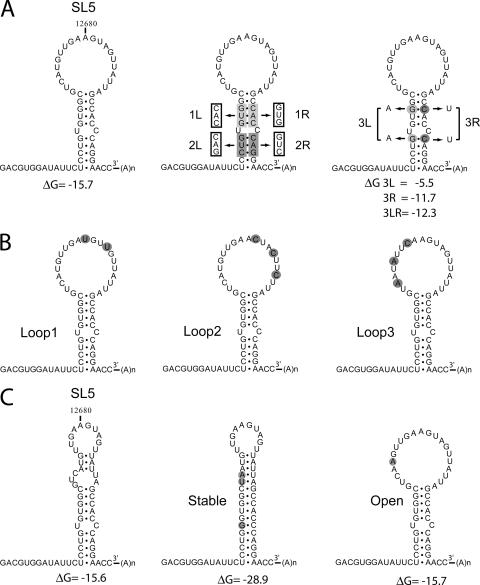
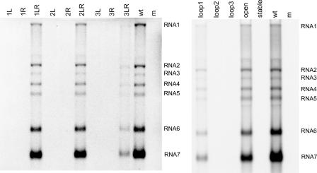
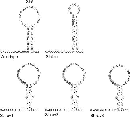
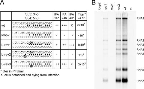
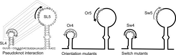
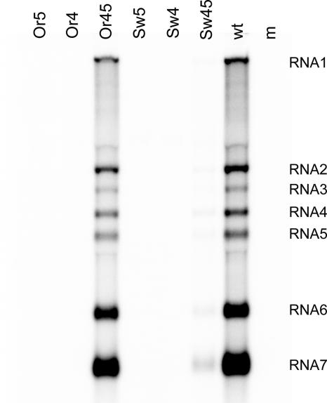
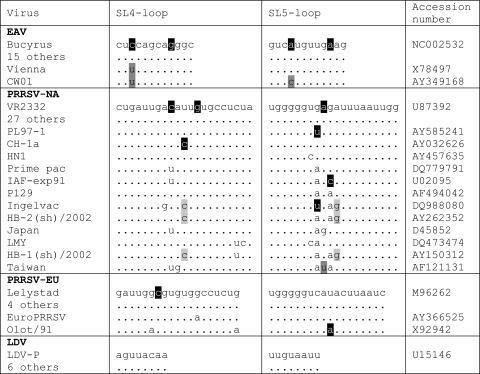
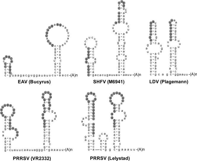
In the life cycle of plus-strand RNA viruses, the genome initially serves as the template for both translation of the viral replicase gene and synthesis of minus-strand RNA and is ultimately packaged into progeny virions. These various processes must be properly balanced to ensure efficient viral proliferation. To achieve this, higher-order RNA structures near the termini of a variety of RNA virus genomes are thought to play a key role in regulating the specificity and efficiency of viral RNA synthesis. In this study, we have analyzed the signals for minus-strand RNA synthesis in the prototype of the arterivirus family, equine arteritis virus (EAV). Using site-directed mutagenesis and an EAV reverse genetics system, we have demonstrated that a stem-loop structure near the 3' terminus of the EAV genome is required for RNA synthesis. We have also obtained evidence for an essential pseudoknot interaction between the loop region of this stem-loop structure and an upstream hairpin residing in the gene encoding the nucleocapsid protein. We propose that the formation of this pseudoknot interaction may constitute a molecular switch that could regulate the specificity or timing of viral RNA synthesis. This hypothesis is supported by the fact that phylogenetic analysis predicted the formation of similar pseudoknot interactions near the 3' end of all known arterivirus genomes, suggesting that this interaction has been conserved in evolution.


Figures

FIG. 1.
FIG. 1.
RNA secondary structure models for the 3′ UTR of wild-type EAV and SL5 mutants. (A) Mutations were introduced in the SL5 stem region. Mutant 1L has a 3-nucleotide substitution on the left side of the stem, and mutant 1R on the right side. Mutations 1L and 1R are complementary, and base pairing is restored in the double mutant 1LR. Similar mutations were also introduced in the lower stem segment (2L, 2R, and 2LR). The stability of the stem region was targeted in mutants 3L, 3R, and 3LR by the replacement of two G-C base pairs. (B) Mutants Loop1, Loop2, and Loop3 contain point mutations in the SL5 loop region. (C) Mfold prediction of an alternative structure for the SL5 stem-loop with similar stability, in which part of the loop region is closed by base pairing. This alternative structure was stabilized by the mutations introduced in mutant Stable and prevented by the mutation introduced in mutant Open.
FIG. 2.
FIG. 2.
RNA synthesis by wild-type EAV and SL5 mutants. Infectious RNA was transfected into BHK-21 cells, and intracellular RNA was isolated at 14 h posttransfection. The RNA was separated in a denaturing agarose gel and analyzed by hybridization to an oligonucleotide detecting all plus-stranded viral RNAs. The positions of the genome (RNA1) and sg mRNAs (RNA2 to RNA7) are indicated.
FIG. 3.
FIG. 3.
Reversion of mutant Stable. The mutations originally introduced into mutant Stable are boxed. Upon sequence analysis, we identified two clones named St-rev1 that contained an insertion partly restoring the wild-type sequence and opening the loop region (insertions are shown in balls, wild-type residues are marked in light gray, mutant residues in dark gray). The sequence St-rev2 was obtained from one clone, the sequence St-rev3 from four clones. After one additional virus passage, all eight clones sequenced were of the St-rev3 type.
FIG. 4.
FIG. 4.
Reversion of mutant Loop2. (A) Sequences of the 3′ terminal region of the genome of the Loop2 revertant were determined by RT-PCR, followed by sequencing of nine individual clones. Of the sequenced clones, seven were found to contain a second-site mutation in the SL5 loop region (L-rev1), but two clones acquired mutations in the SL4 domain. The revertant sequences were introduced into the wild-type EAV cDNA clone. Virus replication was studied using IFA with EAV-specific antisera for nsp3 and N at different time points after transfection, and virus titration was performed using plaque assays. The sequence of the SL5 loop region is depicted in the 3′-to-5′ direction, whereas that of SL4 is depicted in the 5′-to-3′ direction. Base-pairing possibilities between the two sequences are indicated by dots. The mutations introduced in the Loop2 mutant are marked in italics; the acquired reversions are marked by a black box. (B) Hybridization analysis of the RNA synthesis of the loop2 revertants (see the legend to Fig. 2 for details).
FIG. 5.
FIG. 5.
RNA secondary structure model showing the proposed SL4-SL5 pseudoknot interaction and pseudoknot mutants. The nucleotides involved in the pseudoknot interaction are marked in gray, and the base-pairing interaction is depicted by lines. The orientation of the central pentanucleotide in the loop was changed in SL4 (Or4) or SL5 (Or5) or both (Or45), which restored base-pairing possibilities. The pentanucleotide sequence of SL4 was switched with that of SL5 in mutant Sw4, and vice versa in mutant Sw5. Base-pairing possibilities were again restored in mutant Sw45. The stop codon of the N protein gene is underlined.
FIG. 6.
FIG. 6.
Hybridization analysis of the RNA synthesis by wild-type EAV and pseudoknot mutants (see the legend to Fig. 2 for details).
FIG. 7.
FIG. 7.
Alignment of the nucleotides involved in the pseudoknot interaction in different arteriviruses. Several nucleotide changes were identified, as follows: those not disrupting base pairing (unmarked) and disrupting base pairing (black), those creating an additional base pair (dark gray), and covariations (light gray).
FIG. 8.
FIG. 8.
Conservation of the pseudoknot interaction in all known arteriviruses. The RNA secondary structure predictions for the two terminal stem-loop structures in different arterivirus genomes are shown. For all arteriviruses, a putative interaction between the top region of the terminal hairpin and the upstream hairpin is predicted and is marked in gray. Shown are EAV (Brucyrus [accession number NC002532]), SHFV (M6941 [accession number NC003092]), LDV (Plagemann [accession number U15146]), PRRSV (VR2332 [accession number U87392]), and PRRSV (Lelystad [accession number M96262]).

Similar articles

Cited by

References

    1. Almazan, F., C. Galan, and L. Enjuanes. 2004. The nucleoprotein is required for efficient coronavirus genome replication. J. Virol. 78:12683-12688. - PMC - PubMed
    1. Baric, R. S., G. W. Nelson, J. O. Fleming, R. J. Deans, J. G. Keck, N. Casteel, and S. A. Stohlman. 1988. Interactions between coronavirus nucleocapsid protein and viral RNAs: implications for viral transcription. J. Virol. 62:4280-4287. - PMC - PubMed
    1. Beerens, N., and E. J. Snijder. 2006. RNA signals in the 3′ terminus of the genome of equine arteritis virus are required for viral RNA synthesis. J. Gen. Virol. 87:1977-1983. - PubMed
    1. Brierley, I., P. Digard, and S. C. Inglis. 1989. Characterization of an efficient coronavirus ribosomal frameshifting signal: requirement for an RNA pseudoknot. Cell 57:537-547. - PMC - PubMed
    1. den Boon, J. A., E. J. Snijder, E. D. Chirnside, A. A. de Vries, M. C. Horzinek, and W. J. M. Spaan. 1991. Equine arteritis virus is not a togavirus but belongs to the coronaviruslike superfamily. J. Virol. 65:2910-2920. - PMC - PubMed

Publication types

LinkOut - more resources