. 2007 Sep;81(17):9451-60.
doi: 10.1128/JVI.00499-07. Epub 2007 Jun 20.

Arenavirus Z-glycoprotein association requires Z myristoylation but not functional RING or late domains

Affiliations

Arenavirus Z-glycoprotein association requires Z myristoylation but not functional RING or late domains

Althea A Capul et al. J Virol. 2007 Sep.

Abstract

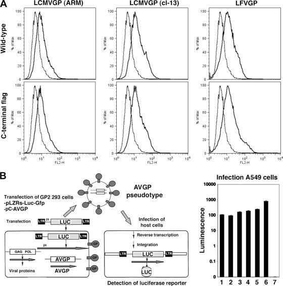
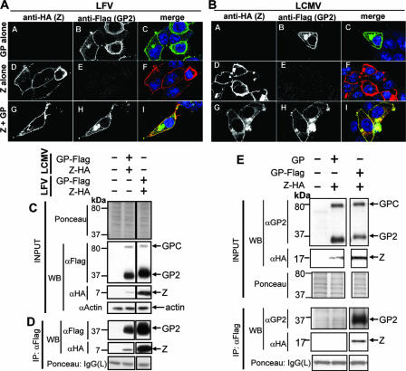
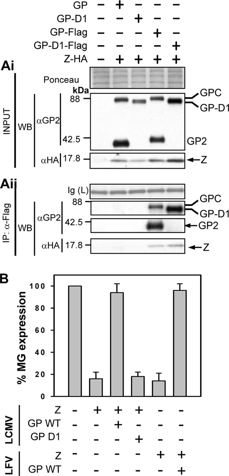
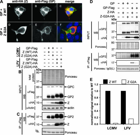
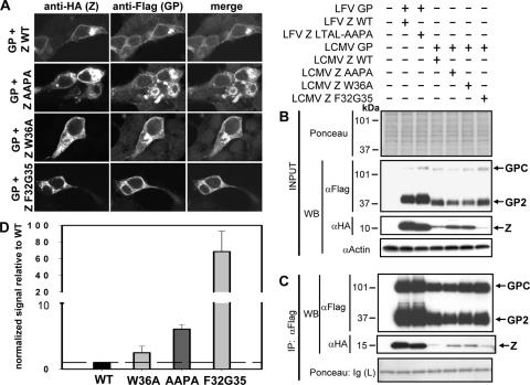
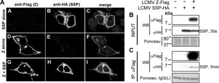
Generation of infectious arenavirus-like particles requires the virus RING finger Z protein and surface glycoprotein precursor (GPC) and the correct processing of GPC into GP1, GP2, and a stable signal peptide (SSP). Z is the driving force of arenavirus budding, whereas the GP complex (GPc), consisting of hetero-oligomers of SSP, GP1, and GP2, forms the viral envelope spikes that mediate receptor recognition and cell entry. Based on the roles played by Z and GP in the arenavirus life cycle, we hypothesized that Z and the GPc should interact in a manner required for virion formation. Here, using confocal microscopy and coimmunoprecipitation assays, we provide evidence for subcellular colocalization and biochemical interaction, respectively, of Z and the GPc. Our results from mutation-function analysis reveal that Z myristoylation, but not the Z late (L) or RING domain, is required for Z-GPc interaction. Moreover, Z interacted directly with SSP in the absence of other components of the GPc. We obtained similar results with Z and GPC from the prototypical arenavirus lymphocytic choriomeningitis virus and the hemorrhagic fever arenavirus Lassa fever virus.


Figures

FIG. 1.
FIG. 1.
Functional characterization of GP-Flag. (A) Surface localization of GP is unaltered by the presence of the C-terminal Flag tag in GP. 293T cells were transfected with plasmids containing LCMV (ARM) GP, LCMV (clone 13) GP, LFV (Josiah) GP, or their Flag-tagged counterparts, and surface GP was determined using FACS analysis. Fluorescence intensity is shown on the x axis, and relative cell counts on the y axis. (B) Murine leukemia virus (MLV) virions pseudotyped with arenavirus GP-Flag show infection efficiencies similar to those with untagged GP pseudotypes. The arenavirus GP constructs from panel A were used to prepare MLV pseudotypes (AVGP) and infect A549 cells following the schematic. Luciferase activity of the various pseudotypes is shown in the bar graph. Lanes: 1, LCMV ARM GP-Flag; 2, LCMV GP cl-13-Flag; 3, LFV GP-Flag; 4, LCMV ARM GP; 5, LFV GP; 6, VSV GP; 7, no GP.
FIG. 2.
FIG. 2.
Subcellular localization and biochemical association of Z and GP. (A) LFV Z and GP colocalize in transfected cells. 293T cells were transiently transfected with LFV Z-HA, GP-Flag, or both and subjected to indirect immunofluorescence. Panels A A, A B, and A C correspond to cells transfected with LFV GP-Flag only; while panels D, E, and F correspond to cells transfected with LFV Z-HA only. Z-HA or GP-Flag was detected using anti-HA (mouse) and anti-Flag (rabbit) antibodies simultaneously to assess cross-reactivity and background. Panels A G to A I and B G to I correspond to cells expressing both Z-HA and GP-Flag, which show colocalization of both proteins. (B) LCMV Z and GP colocalize in transfected cells. Panel B is arranged as in panel A. Results are representative of several independent experiments. (C) Expression of GP and Z proteins. 293T cells were transiently transfected to express Z-HA and GP-Flag, as indicated above the lanes, and total cell lysates were subjected to Western analysis. As a negative control, cells were transfected with empty vector. A section of the membrane stained with Ponceau-S is shown to reflect equal loading. It was noted that expression of LCMV Z-HA was less than that of LFV Z-HA despite the use of the same plasmid to express these proteins. (D) Z and GP biochemically associate. Cell lysates shown in panel C were immunoprecipitated using an anti-Flag antibody, and precipitated proteins were subjected to Western analysis. We could readily detect Z-HA from both LCMV and LFV in Flag immunoprecipitates containing the homologous GPs. (E) Z association with GP is specific. As a control to assess nonspecific interactions, cells were transfected with Z-HA and untagged GP and then subjected to anti-Flag immunoprecipitation. Lanes unrelated to this experiment were cropped. Z-HA was undetectable in immunoprecipitates from cells coexpressing untagged GP, while it was enriched in immunoprecipitates after coexpression with GP-Flag.
FIG. 3.
FIG. 3.
Biochemical and genetic interactions of Z and GP. (Ai) Expression of the LCMV Z-HA, the GP WT, the GP-D1 mutant, and Flag-tagged GPs in total cell lysates. The various GPs were detected using an anti-GP2 antibody, and Z was detected using an anti-HA antibody. A section of the membrane stained with Ponceau-S is shown to reflect equal loading. (Aii) The GP-D1 mutant associates biochemically with Z. Cell lysates shown in panel Ai were immunoprecipitated using an anti-Flag antibody, and the precipitated proteins were analyzed by Western blotting using an antibody to GP2. Z was visualized by Western blotting with anti-HA as shown in the panel, and bands of interest are indicated with arrows. As controls, cells were transfected with empty vector and with the WT GP or the GP-D1 mutant in the presence of Z-HA and then subjected to anti-Flag IP, which allows detection of nonspecific IP with the anti-Flag resin. To show equal loading, the IgG(L) fragment that stains clearly with Ponceau-S on the membrane is shown. (B) Z-mediated inhibition of the LCMV MG is prevented by the WT GP but not by the GP-D1 mutant. Cells (293T) were transfected with the indicated combination of plasmids, and 72-h cell lysates were prepared for CAT assays. CAT activities were normalized by assigning 100% to the activity determined in lysates from cells transfected with L + NP + T7RP + MG and after subtracting background levels of CAT activity detected in lysates of transfected cells that did not receive the plasmid encoding the L polymerase.
FIG. 4.
FIG. 4.
Myristoylation greatly impacts the level of Z-GP association. (A) The Z-G2A mutant shows altered subcellular localization. LCMV Z-G2A HA and GP-Flag localization was assessed in transfected cells by using indirect immunofluorescence. As a positive control, indirect immunofluorescence of cells transfected with Z-HA WT and GP-Flag is shown. Panels A A and A D correspond to signals for Z only, panels A B and A E correspond to signals for GP only, while panels A C and A F show a merged image of the Z, GP, and nuclear signals. (B) Expression of GP-Flag and Z-G2A HA proteins. Cells were transfected with empty vector as a negative control and with GP-Flag and Z WT as a positive control, and total cell lysates were subjected to Western analysis. The lanes are labeled above panel B according to the Z-+-GP combination expressed in the transfectants. Staining of the membrane with Ponceau-S and probing for beta-actin were done to assess loading. It was noted that more Z-G2A was detectable than Z WT in cells. (C) The Z-G2A myristoylation mutant is decreased in biochemical association to GP. Cell lysates shown in panel B were subjected to anti-Flag immunoprecipitation. As controls to assess nonspecific associations, cells were cotransfected with the Z WT or Z-G2A and untagged GP. The amount of Z-G2A detectable in immunoprecipitates containing GP-Flag was similar to that which was detectable in the presence of untagged GP. (D) Densitometry analysis of the coimmunoprecipitated Z-G2A shown in panel B. The amount of Z-G2A was normalized to both the amount of the WT Z and the amount of actin in total cell protein. Results are representative of at least two independent experiments.
FIG. 5.
FIG. 5.
Late and RING domains are not required for Z-GP association. (A) Subcellular localization of Z containing mutations in the late and RING domains. Cells were cotransfected with GP-Flag and various Z mutants, each containing a C-terminal HA tag, and subjected to indirect immunofluorescence of GP and Z. Images of a cell cotransfected to express Z WT and GP are shown as a control. We observed no change in the localization of the Z L domain mutant (Z-AAPA) or the Z-A36 mutant and their colocalization with GP; however, we observed a vesicular pattern in cells expressing the Z F32G35 mutant. (B) Expression of Z mutants in total cell protein. Transiently transfected 293T cells with the GP-Z combinations described in the legend to panel A were lysed, and proteins were detected by Western analysis using anti-Flag and anti-HA antibodies. To assess loading, the membrane was stained with Ponceau-S before probing for GP and Z and also probed for actin specifically. The amounts of GP expression appeared equal among the transfectants, while LCMV Z-AAPA and LCMV Z-A36 showed slightly more expression than LCMV Z WT. There were no differences in expression between LFV Z WT and LFV Z-AAPA-LTAL, but we consistently observed less Z-F32G35 in the βOG-soluble fraction of cells coexpressing GP. Densitometry analysis was used to normalize the expression of Z mutants to that of WT. (C) Late and RING domains are not required to mediate interaction of Z with GP. The cell lysates shown in panel B were subjected to Flag immunoprecipitation and Western blotting to detect Z WT strains and Z mutants. No differences were observed between LFV Z WT and LFV Z-AAPA-LTAL in Z-GP association, and the amounts of LCMV Z WT, LCMV Z-AAPA, and LCMV Z-A36 were consistent with levels in total cell protein. We observed more LCMV Z-F32G35 than LCMV Z WT in immunoprecipitates, however. (D) Densitometry analysis of Z L and RING domain mutants following Flag immunoprecipitation. A separate experiment containing duplicate transfections of the Z L and RING domain mutants was performed to assess the differences in association between the various Z mutants and GP shown in panel C. Following normalization of Z expression in whole-cell lysates, the amount of Z mutants present in Flag immunoprecipitates was compared to that of Z WT. This analysis revealed a modest increase in coimmunoprecipitation of Z-AAPA and Z-A36 with GP and a large increase in Z-F32G35 present in GP immunoprecipitates.
FIG. 6.
FIG. 6.
Subcellular localization and biochemical association between LCMV Z and SSP. (A) LCMV Z and SSP minimally colocalize. 293T cells expressing LCMV Z-Flag and SSP-HA in the indicated combinations were subjected to indirect immunofluorescence and confocal laser scanning microscopy. As described in the legend to Fig. 2, cells expressing Z-Flag or SSP-HA alone were probed simultaneously with anti-Flag or anti-HA antibodies to assess cross-reactivity and background. We observed modest levels of colocalization of Z and SSP in cotransfected cells, which were 25% and 23% of Z with SSP and the reverse, respectively, as determined with the LSM Image Examiner program to calculate the Pearson product-moment correlation coefficient for the respective signals of Z and SSP. (B) Expression of LCMV Z-Flag and SSP-HA in total cell protein. 293T cells were transiently transfected with LCMV Z-Flag and LCMV SSP-HA, and both proteins were detected in cells by Western analysis of whole-cell lysates. To observe loading, the membrane was stained with Ponceau-S before probing with antibodies, and the length of exposure time following enhanced chemiluminescence to detect SSP-HA is noted. (C) LCMV Z and SSP biochemically associate. Western analysis of anti-Flag immunoprecipitates was done for SSP-HA following coexpression with LCMV Z-Flag. As a control for specificity, anti-Flag immunoprecipitation was done following expression of SSP-HA alone in cells. Densitometry analysis of these exposures indicated that the specific co-IP signal from SSP-HA with LCMV Z-Flag was 9.7-fold higher than the condition where SSP-HA was expressed alone.

References

    1. Agnihothram, S. S., J. York, and J. H. Nunberg. 2006. Role of the stable signal peptide and cytoplasmic domain of G2 in regulating intracellular transport of the Junin virus envelope glycoprotein complex. J. Virol. 80:5189-5198. - PMC - PubMed
    1. Aizaki, H., K. J. Lee, V. M. Sung, H. Ishiko, and M. M. Lai. 2004. Characterization of the hepatitis C virus RNA replication complex associated with lipid rafts. Virology 324:450-461. - PubMed
    1. Barton, L. L., M. B. Mets, and C. L. Beauchamp. 2002. Lymphocytic choriomeningitis virus: emerging fetal teratogen. Am. J. Obstet. Gynecol. 187:1715-1716. - PubMed
    1. Bavari, S., C. M. Bosio, E. Wiegand, G. Ruthel, A. B. Will, T. W. Geisbert, M. Hevey, C. Schmaljohn, A. Schmaljohn, and M. J. Aman. 2002. Lipid raft microdomains: a gateway for compartmentalized trafficking of Ebola and Marburg viruses. J. Exp. Med. 195:593-602. - PMC - PubMed
    1. Bergthaler, A., N. U. Gerber, D. Merkler, E. Horvath, J. C. de la Torre, and D. D. Pinschewer. 2006. Envelope exchange for the generation of live-attenuated arenavirus vaccines. PLoS Pathog. 2:e51. - PMC - PubMed

Publication types

MeSH terms

Substances

LinkOut - more resources