. 2007 Jun 21;54(6):919-31.
doi: 10.1016/j.neuron.2007.05.029.

Activity-dependent validation of excitatory versus inhibitory synapses by neuroligin-1 versus neuroligin-2

Affiliations

Activity-dependent validation of excitatory versus inhibitory synapses by neuroligin-1 versus neuroligin-2

Alexander A Chubykin et al. Neuron. .

Abstract

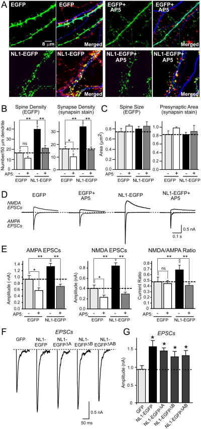
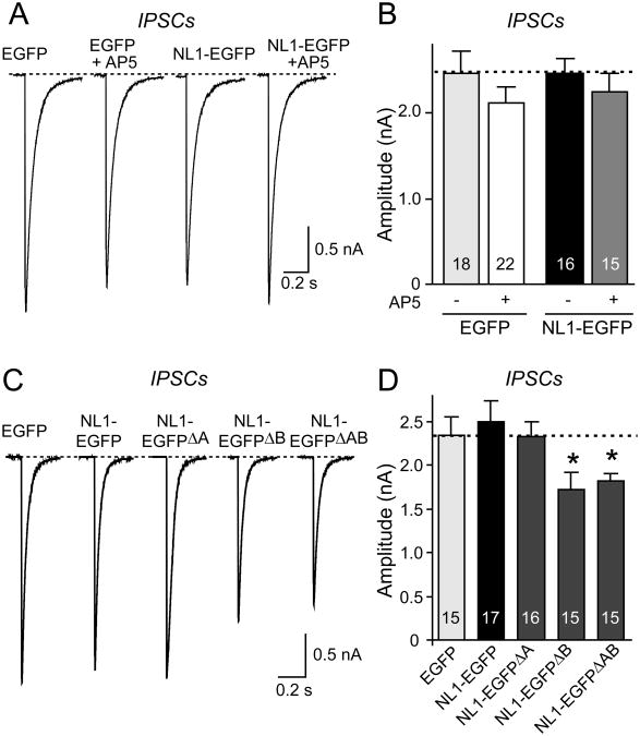
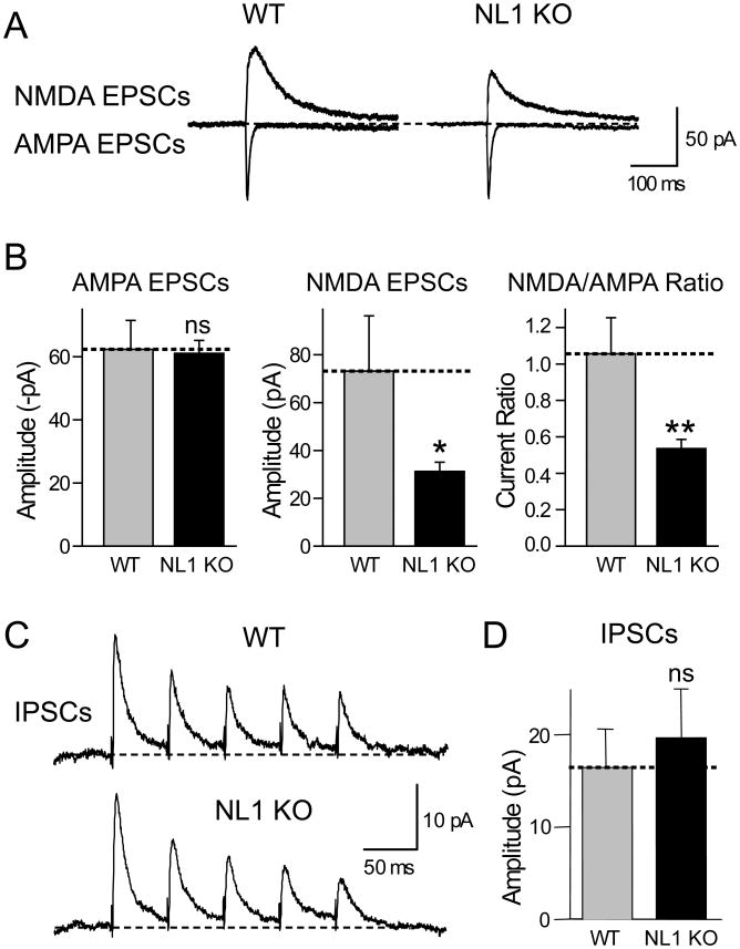
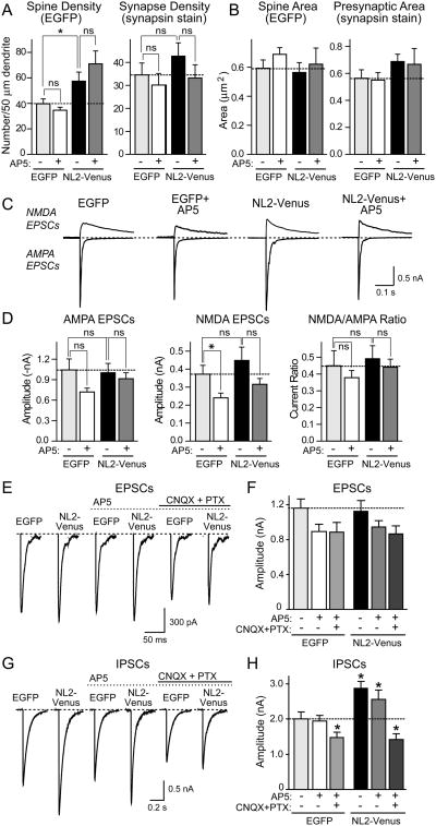
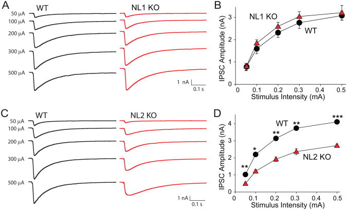
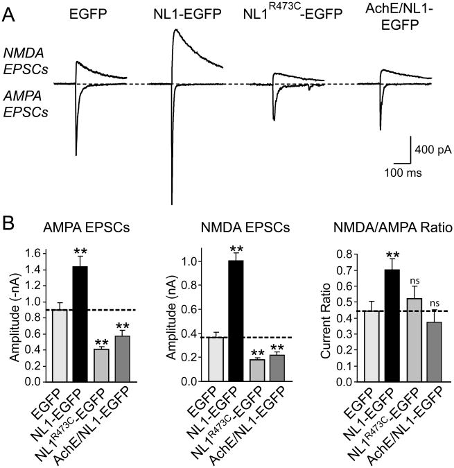
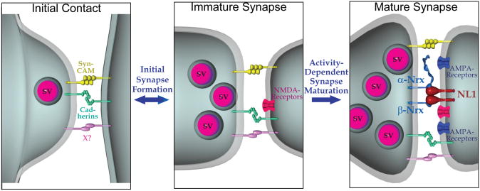
Neuroligins enhance synapse formation in vitro, but surprisingly are not required for the generation of synapses in vivo. We now show that in cultured neurons, neuroligin-1 overexpression increases excitatory, but not inhibitory, synaptic responses, and potentiates synaptic NMDAR/AMPAR ratios. In contrast, neuroligin-2 overexpression increases inhibitory, but not excitatory, synaptic responses. Accordingly, deletion of neuroligin-1 in knockout mice selectively decreases the NMDAR/AMPAR ratio, whereas deletion of neuroligin-2 selectively decreases inhibitory synaptic responses. Strikingly, chronic inhibition of NMDARs or CaM-Kinase II, which signals downstream of NMDARs, suppresses the synapse-boosting activity of neuroligin-1, whereas chronic inhibition of general synaptic activity suppresses the synapse-boosting activity of neuroligin-2. Taken together, these data indicate that neuroligins do not establish, but specify and validate, synapses via an activity-dependent mechanism, with different neuroligins acting on distinct types of synapses. This hypothesis reconciles the overexpression and knockout phenotypes and suggests that neuroligins contribute to the use-dependent formation of neural circuits.


Figures

Figure 1
Figure 1. Chronic NMDA-receptor block suppresses NL1-induced increase in the number of functional excitatory synapses
(A) Representative images of hippocampal neurons transfected with NL1-EGFP or EGFP alone, and cultured in the presence or absence of 50 μM AP5 for four days. Neurons were visualized by EGFP fluorescence (green), and immunolabeling with antibodies to the dendritic marker MAP2 (blue) and the presynaptic marker synapsin (red). For each sample, the EGFP image is shown on the left, whereas the merged image for EGFP, MAP2, and synapsin is shown on the right. (B) and (C) Quantitative analyses of synapse numbers (B) and size (C) in neurons expressing EGFP or EGFP-tagged NL1, and treated with either control medium or AP5. For an analysis of specifically GABAergic synapses, see Suppl. Fig. 2. (D) Representative electrophysiological recordings of evoked NMDA- and AMPA-receptor dependent EPSCs in neurons transfected with EGFP or NL1-EGFP with or without NMDA-receptor blockade by AP5. Recordings were made in the absence of AP5. (E) Amplitudes of AMPA- and NMDA-receptor dependent EPSCs and NMDA/AMPA ratio in neurons transfected with EGFP or NL1-EGFP with and without chronic AP5 treatment. (F) and (G) Representative traces (F) and summary graphs (G) of electrophysiological recordings of AMPA-dependent EPSCs in neurons transfected with control vector or vectors expressing all four alternative splice variants of NL1 (Boucard et al., 2005). Data shown in (B), (C), (E), and (G) are means ± SEMs (n=3 independent experiments with 6-10 neurons/experiment and condition); asterisks indicate statistically significant differences (* = p<0.05; ** = p<0.01; ns = not significant). In all experiments in this and all following figures, the NL splice variant analyzed corresponds to the variant with inserts in all sites of alternative splicing except when indicated otherwise.
Figure 2
Figure 2. NL1 expression does not alter IPSCs: Effect of chronic NMDA-receptor blockade and alternative splicing
Sample traces (A) and summary graphs (B) of IPSCs recorded from neurons expressing only EGFP or EGFP-tagged NL1 cultured either in control medium or in medium containing 50 μM AP5 for four days prior to the recordings. (C) and (D) Alternative splicing of NL1 does not activate its lack of an effect on IPSCs. IPSCs were monitored in 50 μM AP5 and 10 μM CNQX (means ± SEMs; n=18 cells/3 cultures; asterisks represent statistically significant difference: ** =p<0.01; ns, not significantly different).
Figure 3
Figure 3. Chronic blockade of CaM-Kinase IIa mimics the effect of NMDA-receptor blockade on EPSCs in neurons expressing NL1
Sample traces (A) and summary graphs (B) of EPSCs recorded from neurons expressing only EGFP or EGFP-tagged NL1 cultured either in medium containing 5 μM KN-93 ( CaM-Kinase IIα inhibitor) or in control medium containing 5 μM KN-92 ( inactive analog of KN-93) for four days prior to the recordings. Data shown are means ± SEMs; asterisks represent statistically significant differences (n=18 cells/3 cultures; *=p<0.05, ** =p<0.01; ns, not significantly different).
Figure 4
Figure 4. Deletion of NL1 in KO mice lowers the NMDA/AMPA-receptor ratio without significantly altering the IPSC amplitude
(A) Representative traces of NMDA- (top) and AMPA-receptor dependent EPSCs (bottom) evoked by local stimulation with a microelectrode and recorded from a pyramidal neuron in the CA1 region of the hippocampus from littermate wild-type and NL1 KO mice. (B) Mean amplitudes of NMDA- and AMPA-receptor dependent EPSCs and mean NMDA/AMPA-receptor dependent EPSC ratio. Stimulation strength was adjusted to yield similar AMPA-receptor dependent EPSC amplitudes, and NMDA-receptor dependent EPSCs were then measured in the same neuron with the same stimulus strength (n=12 for each genotype). (C) and (D) Representative traces (C) and mean amplitudes of IPSCs (D) monitored by paired recordings from adjacent inhibitory and excitatory neurons in the somatosensory cortex (n=12 wild-type and n=10 NL1 KO neurons; data shown are means ± SEMs; asterisks indicate if there is a statistically significant difference between WT and NL1 KO, * = p<0.05; ** = p<0.01).
Figure 5
Figure 5. NL2 selectively enhances inhibitory synaptic function
Hippocampal neurons were transfected with NL2-Venus or EGFP, and cultured in the presence or absence of 50 μM AP5 for four days. (A) and (B) Summary graphs of the quantitative analysis of synapse numbers (A) and size (B) in neurons expressing EGFP or NL2-Venus, and treated with either control medium or AP5. For representative images, see Suppl. Fig. 3. (C) Representative electrophysiological traces of evoked NMDA- and AMPA-receptor dependent EPSCs in neurons transfected with EGFP or NL2-Venus with or without NMDA-receptor blockade. (D) Amplitudes of AMPA- and NMDA-receptor dependent EPSCs and the NMDA/AMPA ratio in neurons transfected with EGFP and NL2-Venus with and without chronic AP5 treatment. (E) - (H) Effect of chronic treatments with AP5 without and with CNQX and picrotoxin on evoked EPSCs (E and F) and IPSCs (G and H) in NL2-overexpressing neurons. Panels show sample traces (E and G) and summary graphs (F and H). Neurons were transfected at 10 DIV and incubated in 50 μM AP5 with or without 10 μM CNQX and 50 μM picrotoxin for 4 days. IPSCs were monitored in 50 μM AP5 and 10 μM CNQX. Data shown in (A), (B), (D), (F) and (H) are means ± SEMs (n=3 independent experiments with 6-10 neurons/experiment and condition); asterisks indicate statistically significant differences (* = p<0.05; ** = p<0.01; ns - not significant).
Figure 6
Figure 6. Deletion of NL2 but not of NL1 KO depresses IPSC amplitudes in acute cortical slices
Evoked IPSCs were measured as a function of the stimulus strength in Layer 2/3 of the somatosensory (barrel) cortex in response to extracellular stimulation by a microelectrode positioned nearby. (A) and (B), and (C) and (D) show representative traces (A, C) and summary graph (B, D) for evoked IPSCs from NL1 (A, B) and NL2 KO mice (C,D), respectively, and their wild-type littermate controls (n=4 mouse pairs each). Data shown in (B) and (D) are means ± SEMs; asterisks indicate statistically significant differences (* = p<0.05; ** = p<0.01; *** = p<0.001).
Figure 7
Figure 7. Autism mutation of NL1 causes dominant-negative suppression of EPSCs
(A) Representative traces of NMDA- (top) and AMPA-receptor dependent EPSCs (bottom). For a diagram of the mutants and a morphological analysis of the effect of the mutants on synapse density, see Suppl. Fig. 4. (B) Mean amplitudes of NMDA- and AMPA-receptor dependent EPSCs and mean NMDA/AMPA receptor ratios. Data shown are means ± SEMs (n=18 neurons from 3 cultures); asterisks indicate that a condition exhibits a statistically significant difference from the EGFP-only transfected control condition (* =p<0.05; **=p<0.01).
Figure 8
Figure 8. Model of the role of NL1 in synapse formation
The initial synaptic contact between neurons is proposed to involve multiple cell-adhesion molecules, including SynCAM and cadherins which might impart specificity on synaptic contacts. The resulting immature synapses are functional, but are stabilized and further specified in terms of their specific properties (release probability, plasticity, NMDA/AMPA-receptor ratio, and others) by activity-dependent processes. The model suggests that NL1 mediates the activity-dependent stabilization of transient synaptic contacts, but that this function of NL1 depends on the simultaneous activation of NMDA-receptors. In promoting activity-dependent synapse stabilization, postsynaptic NL1 likely transduces a trans-synaptic signal triggered by binding of its extracellular esterase-like domain to presynaptic neurexins. NL2 presumably performs an analogous function in inhibitory synapses.

References

    1. Agmon A, Connors BW. Thalamocortical response of mouse somatosensory (barrel) cortex in vitro. Neuroscience. 1991;41:365–379. - PubMed
    1. Alvarez VA, Ridenour DA, Sabatini BL. Retraction of synapses and dendritic spines induced by off-target effects of RNA interference. J Neurosci. 2006;26:7820–7825. - PMC - PubMed
    1. Biederer T, Sara Y, Mozhayeva M, Atasoy D, Liu X, Kavalali ET, Sudhof TC. SynCAM, a synaptic adhesion molecule that drives synapse assembly. Science. 2002;297:1525–1531. - PubMed
    1. Boucard AA, Chubykin AA, Comoletti D, Taylor P, Sudhof TC. A Splice Code for trans-Synaptic Cell Adhesion Mediated by Binding of NL1 to alpha- and beta-Neurexins. Neuron. 2005;48:229–236. - PubMed
    1. Cantallops I, Cline HT. Synapse formation: if it looks like a duck and quacks like a duck. Curr Biol. 2000;10:R620–623. - PubMed

Publication types

MeSH terms