. 2007 Jun 21;54(6):949-59.
doi: 10.1016/j.neuron.2007.06.002.

Differential expression of posttetanic potentiation and retrograde signaling mediate target-dependent short-term synaptic plasticity

Affiliations

Differential expression of posttetanic potentiation and retrograde signaling mediate target-dependent short-term synaptic plasticity

Michael Beierlein et al. Neuron. .

Abstract

Short-term synaptic plasticity influences how presynaptic spike patterns control the firing of postsynaptic targets. Here we investigated whether specific mechanisms of short-term plasticity are regulated in a target-dependent manner by comparing synapses made by cerebellar granule cell parallel fibers onto Golgi cells (PF-->GC synapse) and Purkinje cells (PF-->PC synapse). Both synapses exhibited similar facilitation, suggesting that any differential short-term plasticity does not reflect differences in the initial release probability. PF-->PC synapses were highly sensitive to stimulus bursts, which could result in either depression of subsequent responses, mediated by endocannabinoid-dependent retrograde signaling, or enhancement of responses through posttetanic potentiation (PTP). In contrast, stimulus bursts had remarkably little effect on the strength of PF-->GC synapses. Unlike PCs, GCs were unable to regulate their PF synapses by releasing endocannabinoids. Moreover, PTP was reduced at the PF-->GC synapse compared to the PF-->PC synapse. Thus, the target-dependence of PF synapses arises from the differential expression of both retrograde signaling and PTP.


Figures
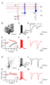
Figure 1
Figure 1. Target cell-specific short-term enhancement and suppression at parallel fiber synapses
(A) A simplified circuit of the cerebellar cortex shows excitatory synapses made by granule cell (GrC) parallel fibers (PFs) onto Purkinje cells (PCs) and Golgi cells (GCs). Arrows indicate that mossy fibers (MFs) provide a major source of excitatory input to the cerebellar cortex and PCs are the sole output from this region. ML, molecular layer; PL, Purkinje cell layer; GrL, granular layer. (B, H) Fluorescent images of a PC (B) and a GC (H) filled with Alexa 594. Postsynaptic responses to PF burst stimulation (10 stimuli, 50 Hz) were recorded in current clamp (C, F, I, L). PFs were activated with an extracellular electrode placed either in the ML (C, D, I, J, black traces) or the GrL, (F, G, L, M, red traces). EPSCs evoked at 0.5 Hz prior to and following a PF burst were recorded in voltage-clamp, at a holding potential of −70 mV in a representative PC (D, G) and a GC (J, M), using a K-based internal solution. (E, K) Time course of change in synaptic strength following PF bursts (at t = 0 s) evoked by ML (black) or GrL stimulation (red). Responses shown are normalized to the average EPSC amplitude prior to burst stimulation.
Figure 2
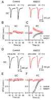
Figure 2. Target cell-dependent short-term suppression is mediated by endocannabinoids
Synaptically-evoked suppression of excitation (SSE; A, B) and depolarization-induced suppression of excitation (DSE; D, E) were examined at PF→GC synapses. Summary data of PC SSE (C) and DSE (F) are shown for comparison. Experiments were performed in control conditions (black traces and black symbols) and in the presence of the CB1R antagonist AM251 (2 µM, red traces and red symbols). Studies of SSE were performed using a K-based internal solution and PFs were stimulated in the ML. Studies examining DSE were performed with a Cs-based internal solution. (A) EPSCs recorded from a GC, evoked prior to (pre-burst) and 3 s following PF burst stimulation. Time course of EPSC amplitudes prior to and following burst stimulation is shown for synapses onto GCs (B; n = 5 cells in control, n = 5 cells in AM251) and PCs (C; n = 6 cells in control, n = 5 cells in AM251). (D–F) Cells were depolarized to 0 mV for 2 s. Representative recordings are shown for a GC (D) and the time course of synaptic strength prior to and following postsynaptic depolarization is shown for GCs (E; n = 15 cells in control, n = 7 cells in AM251) and PCs (F; n = 7 cells in control, n = 6 cells in AM251). Figures 2C and 2F reproduced from (Beierlein and Regehr, 2006) to allow comparison.
Figure 3
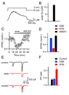
Figure 3. Calcium signals in GC dendrites and presynaptic expression of CB1R at PF to GC synapses
(A) A representative experiment is shown in which a GC was voltage-clamped, depolarized from −70 mV to 0 mV for 2 s, and the resulting dendritic calcium signal was quantified with Fura-FF. (B) Average peak dendritic calcium levels measured in 6 GCs. (C) Bath application of the CB1R agonist WIN 55,212-2 (WIN, 2 µM) reduces the amplitude of PF→GC EPSCs. EPSC amplitude is restored following bath application of AM251 (5 µM). (D) Summary of the effects of a 2 s depolarizing step (DSE, blue), WIN (red) and of AM251 (black) on the magnitude of the EPSC in GCs (n = 5). (E–F) Effects of postsynaptic depolarization and WIN on paired-pulse facilitation (20 ms ISI). Experiments were conducted in 4 mM external calcium. As shown for a representative experiment (E), WIN reduced the amplitude of the first and second EPSC. Overlay, with the traces scaled to the amplitude of the first response, shows that WIN increased pairedpulse facilitation. (F) Summary of the average paired-pulse ratio (PPR = EPSC2/EPSC1) in control conditions (black), following a 2 s depolarization (blue), and following WIN application (red, n = 5 GCs).
Figure 4
Figure 4. Target-dependent differences in short-term facilitation and PTP
Paired-pulse facilitation (A, B), enhancement during stimulus trains (C, D), and PTP (E, F) following stimulus trains (10 stimuli at 50 Hz) were examined at PF synapses onto PCs (black) and GCs (red). EPSCs were recorded in voltage-clamp with a Cs-based internal solution in the presence of GABAAR, GABABR, and CB1R antagonists. Summaries of experiments examining each form of plasticity (A, n = 6 PCs and GCs; C, n = 13 PCs and GCs; E, n = 12 PCs and GCs) and representative experiments (B, D, F) are shown.
Figure 5
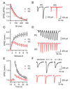
Figure 5. PTP at PF synapses is expressed presynaptically
The magnitude of PTP (A, C) and changes in paired-pulse ratio (EPSC2/EPSC1) during PTP (B, C) were determined following PF bursts (10 stimuli at 50 Hz). (A) Summary of magnitude of PTP, with responses normalized to EPSC1 of the burst (n = 9 PCs, n = 14 GCs). (B) Changes in PPR, for the same data set shown in (A).Values are normalized to PPR at time t = 0 s. Representative experiments (C) are shown for synapses onto PCs (black) and GCs (red).
Figure 6
Figure 6. Target-specific differences in PTP do not depend on initial release probability
(A) Graph plots the magnitude of PTP (EPSC amplitude at Δt = 1 s following burst, normalized to EPSC prior to burst) as a function of paired-pulse ratio for PCs (n = 28, black symbols) and GCs (n = 38, red symbols). Filled symbols denote average values. (B) Average values for PTP and paired-pulse ratio for control conditions (circles, same as (A)), 2 mM DGG (triangles, n = 6 PCs, n = 8 GCs) and 1 mM external calcium (squares, n = 6 PCs, n = 6 GCs), for PCs (black) and GCs (red).
Figure 7
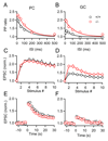
Figure 7. Munc13-3 modulates paired-pulse plasticity without influencing PTP
Short-term plasticity was examined in Munc13-3 knockout mice (red traces) and their wild-type littermates (black traces). Summaries are shown for PPR (EPSC2/EPSC1) measured for a range of inter-stimulus intervals for PCs (A) and GCs (B) (n = 4 to 6 for each condition). Summary data for enhancement during (C, D) and following (E, F) PF bursts (10 stimuli, 50 Hz) (n = 10 to 16 cells for each condition).
Figure 8
Figure 8. PKC is required for PTP
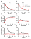
Paired-pulse plasticity (A, B), enhancement during (C, D), and following (E, F) stimulus trains (10 stimuli, 50 Hz) were examined at PF synapses onto PCs and GCs, in control conditions (black symbols) and in the presence of the protein kinase C (PKC) inhibitor GF 109203X (10 µM, red symbols, n = 7 PCs, n = 6 GCs). 3 0

Similar articles

Cited by

References

    1. Abbott LF, Regehr WG. Synaptic computation. Nature. 2004;431:796–803. - PubMed
    1. Alle H, Jonas P, Geiger JR. PTP and LTP at a hippocampal mossy fiber-interneuron synapse. Proc Natl Acad Sci U S A. 2001;98:14708–14713. - PMC - PubMed
    1. Atluri PP, Regehr WG. Determinants of the time course of facilitation at the granule cell to Purkinje cell synapse. J Neurosci. 1996;16:5661–5671. - PMC - PubMed
    1. Augustin I, Betz A, Herrmann C, Jo T, Brose N. Differential expression of two novel Munc13 proteins in rat brain. Biochem J. 1999a;337(Pt 3):363–371. - PMC - PubMed
    1. Augustin I, Korte S, Rickmann M, Kretzschmar HA, Sudhof TC, Herms JW, Brose N. The cerebellum-specific Munc13 isoform Munc13-3 regulates cerebellar synaptic transmission and motor learning in mice. J Neurosci. 2001;21:10–17. - PMC - PubMed

Publication types

MeSH terms

LinkOut - more resources