. 2007 Nov 15;121(10):2153-61.
doi: 10.1002/ijc.22900.

Modulating metastasis by a lymphangiogenic switch in prostate cancer

Affiliations

Modulating metastasis by a lymphangiogenic switch in prostate cancer

Ebba Brakenhielm et al. Int J Cancer. .

Abstract

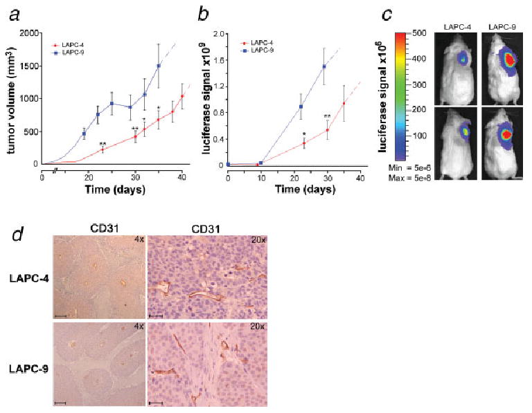
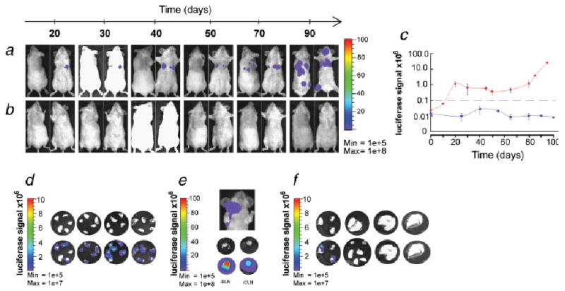
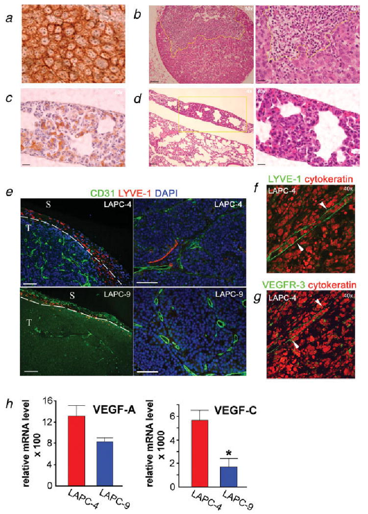
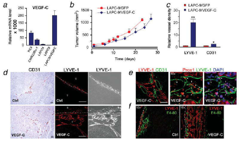
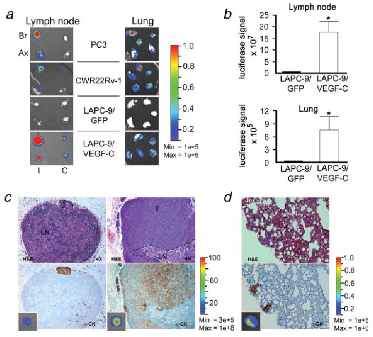
Prostate cancer dissemination is difficult to detect in the clinic, and few treatment options exist for patients with advanced-stage disease. Our aim was to investigate the role of tumor lymphangiogenesis during metastasis. Further, we implemented a noninvasive molecular imaging technique to facilitate the assessment of the metastatic process. The metastatic potentials of several human prostate cancer xenograft models, LAPC-4, LAPC-9, PC3 and CWR22Rv-1 were compared. The cells were labeled with luciferase, a bioluminescence imaging reporter gene, to enable optical imaging. After tumor implantation the animals were examined weekly during several months for the appearance of metastases. Metastatic lesions were confirmed by immunohistochemistry. Additionally, the angiogenic and lymphangiogenic profiles of the tumors were characterized. To confirm the role of lymphangiogenesis in mediating metastasis, the low-metastatic LAPC-9 tumor cells were engineered to overexpress VEGF-C, and the development of metastases was evaluated. Our results show CWR22Rv-1 and PC3 tumor cell lines to be more metastatic than LAPC-4, which in turn disseminates more readily than LAPC-9. The difference in metastatic potential correlated with the endogenous production levels of lymphangiogenic growth factor VEGF-C and the presence of tumor lymphatics. In agreement, induced overexpression of VEGF-C in LAPC-9 enhanced tumor lymphangiogenesis leading to the development of metastatic lesions. Taken together, our studies, based on a molecular imaging approach for semiquantitative detection of micrometastases, point to an important role of tumor lymphatics in the metastatic process of human prostate cancer. In particular, VEGF-C seems to play a key role in prostate cancer metastasis.


Figures

Figure 1
Figure 1
Prostate cancer xenograft models: optical imaging of tumor growth. LAPC-4 and LAPC-9 human tumor cells were implanted in immune-deficient mice (n = 10). Tumor volume was measured (a) and the luciferase reporter gene (FL) activity assayed via in vivo optical imaging (b). Blue color = LAPC-9 group; red color = LAPC-4 group. Dashed extensions of the curves represent projected values after tumors had been surgically removed in the majority of animals in a group. Representative examples of the tumor-derived optical signal in LAPC-4- and LAPC-9-bearing mice at day 20 postimplantation are shown (c). The color bar indicates the intensity range of the bioluminescence signal (p/s/cm2/sr). The tumors were surgically removed within 4–7 weeks postimplantation. Immunohistochemical staining of blood vessels in tumor sections revealed no difference in vascularity between the groups (d; ×4 and ×20). Scale bars = 200 and 50 μm, respectively.
Figure 2
Figure 2
Tumor metastasis monitored by in vivo and ex vivo optical imaging. Representative examples of dorsal and ventral optical images of mice in the LAPC-4- (a) and LAPC-9 cohort (b) at various time points following primary tumor resection are shown. The appearance of bioluminescence signal above background was first evident in the ventral side of the mice, suggestive of lung metastases. The color bar to the right indicates the signal intensity range (p/s/cm2/sr × 106). The maximal luciferase signal emitted from the ventral side of animals in LAPC-4- and LAPC-9 groups is plotted over time after tumor removal (c). The grey dashed line shows the level set for background signal. Only LAPC-4-implanted mice developed in vivo signals indicative of metastasis. These secondary sources of bioluminescence emission in the animals remained stable or increased in intensity over time. Blue color = LAPC-9 group; red color = LAPC-4 group. Data represent averages ±SEM (n = 4). Within 100 days after the tumor removal the animals were sacrificed and the lungs and lymph nodes dissected and imaged optically for the presence of tumor cell-derived luciferase signal. Lung metastatic signs were observed in all but one of the LAPC-4- (n = 9) and in only one of the LAPC-9-implanted mice (n = 7), whereas lymph node signals were found in three LAPC-4 mice and in one LAPC-9 mouse (see Table I). Representative examples of lungs (d) and lymph nodes (e) from the LAPC-4 group and lungs (f) from the LAPC-9 group are shown. The color scale indicates bioluminescence intensity (p/s/cm2/sr). The upper row circular microphotographs are each from a different animal, and the lower row pictures are the corresponding bioluminescence composite images. iBLN, ipsilateral brachial lymph node; iCLN, ipsilateral cervical lymph node.
Figure 3
Figure 3
Immunohistochemical detection of metastases and studies of angiogenic and lymphangiogenic tumor profiles. Lung and lymph node sections from LAPC-4- and LAPC-9-implanted mice were processed to detect tumor cells in the parenchyma. LAPC-4 primary tumor sections served as positive controls for human cytokeratin (a). Hematoxylin & eosin stained sections revealed tumor metastases in the lymph nodes (b) and lungs (d) from the LAPC-4 group. A brachial lymph node from a LAPC-4-implanted mouse shows a large subcapsular lesion (b). Cytokeratin staining revealed tumor cells present in lungs in the LAPC-4-implanted group (c). Yellow lines indicate the tumor mass. Scale bars = 200 μm (×4), 100 μm (×10) and 25 μm (×40), respectively. The blood (CD31; green) and lymph (LYVE-1; red) vessels of the primary tumors (nuclei, blue) were visualized by immunohistochemistry. Analysis of LAPC-4 tumors revealed the presence of small lymphatics both in peritumoral and intratumoral areas (e, left: ×10 and right: ×20; f, ×40). Most LAPC-9 tumors displayed only very few lymphatic vessels at the tumor periphery (e, left: ×10). Scale bar in e = 100 μm. T, tumor; S, surrounding tissue. The intratumoral LYVE-1 positive (green) structures (f, ×40, white arrowheads) were also positive for another lymphatic marker, VEGFR-3 (green) (g, ×40, white arrowheads). Angiogenic and lymphangiogenic growth factor expressions were analyzed in the excised tumors by real time RT-PCR analysis (h). VEGF-A levels were found to be similar whereas VEGF-C expression levels were significantly and markedly elevated in LAPC-4 as compared to LAPC-9 tumors. *p < 0.05.
Figure 4
Figure 4
VEGF-C expression enhances intratumoral lymphangiogenesis in LAPC-9. VEGF-C expression was determined by real time RT-PCR analysis of various prostate cancer tumor samples (a). Overexpression of VEGF-C in LAPC-9 results in > 100-fold increase in VEGF-C levels as compared to LAPC-9/GFP tumors. This is ∼ 2-fold and 4-fold higher levels as compared to PC3 and CWR22Rv-1, respectively. Primary tumor growth rates in immune-deficient mice were similar in LAPC-9/GFP- and LAPC-9/VEGF-C-implanted mice (b; n = 6–8). Tumor immunohistochemical examination revealed that blood vessels (CD31; ×4) and in particular intratumoral lymphatic vessels (LYVE-1, ×20) were more abundant in LAPC-9/VEGF-C tumors as compared to LAPC-9/GFP control tumors (c, d; S, surrounding tissue; T, tumor). Quantification analysis demonstrated that the lymphatic vessel density in LAPC-9/VEGF-C tumors (VEGF-C, blue) was 20-fold higher than in LAPC-9/GFP control tumors (Ctrl, red) (c; LYVE-1), whereas there was only a modest 2-fold increase in blood vessel density (c; CD31). Double staining for lymph (LYVE-1, red) and blood vessels (CD31, green) in LAPC-9/VEGF-C tumors revealed extensive networks of intratumoral lymph vessels growing in proximity to blood vessels (e, left: ×20). LYVE-1 positive structures (green) were also positive for another lymphatic marker, prox1 (red) (e, center and right, ×40, white arrowheads). LYVE-1 positive cells (red) did not colocalize with a macrophage marker F4/80 (green) (f, ×10). White scale bars in d, e = 100 μm; *p < 0.05, **p < 0.005.
Figure 5
Figure 5
VEGF-C expression levels correlate with development of lymph node and lung metastases. Different human prostate cancer cells were implanted in immune-deficient mice (n = 6–8). At sacrifice, the luciferase reporter gene (RL) activity was assayed ex vivo by optical imaging of dissected organs. Representative examples of bioluminescence images of lymph nodes and lungs are shown (a, I, ipsilateral, C, contralateral, Br, brachial lymph node, Ax, axillary lymph node). The color bars indicate the intensity range of the bioluminescence signal (p/s/cm2/sr × 106). The mice bearing PC3 and CWR22Rv-1 tumors showed extensive metastasis to regional lymph nodes and lungs, whereas LAPC-9/GFP control tumors did not metastasize. Overexpression of VEGF-C in LAPC-9 resulted in intense luciferase signals in both lymph nodes and lungs, suggestive of metastasis. Quantification analysis of bioluminescence revealed that the optical signals were ∼ 100-fold higher in regional lymph nodes and ∼ 20-fold higher in lungs from LAPC-9/VEGF-C-implanted mice as compared to LAPC-9-implanted control mice (b). Hematoxylin & eosin (H&E, upper panel) and cytokeratin (α-CK, lower panel) staining of tissue sections confirmed the presence of tumor metastases in the lymph nodes (c; T, tumor; LN, lymph node, ×4) and lungs (d; ×10) from the LAPC-9/VEGF-C-implanted group. The corresponding whole organ optical imaging photo for each sample is shown in the small insert in the lower panels (c, d). The color bars indicate the intensity range of the bioluminescence signal (p/s/cm2/sr × 106). *p < 0.05.

References

    1. National Cancer Institute. Cancer Stat Facts: Surveillance, Epidemiology, and End Results (SEER) Program. Rockville, MD: NCI; 2006.
    1. D'Amico AV, Whittington R, Malkowicz SB, Fondurulia J, Chen MH, Kaplan I, Beard CJ, Tomaszewski JE, Renshaw AA, Wein A, Coleman CN. Pretreatment nomogram for prostate-specific antigen recurrence after radical prostatectomy or external-beam radiation therapy for clinically localized prostate cancer. J Clin Oncol. 1999;17:168–72. - PubMed
    1. Stone NN, Stock RG. Laparoscopic pelvic lymph node dissection in the staging of prostate cancer. Mt Sinai J Med. 1999;66:26–30. - PubMed
    1. Shah RB, Mehra R, Chinnaiyan AM, Shen R, Ghosh D, Zhou M, Macvicar GR, Varambally S, Harwood J, Bismar TA, Kim R, Rubin MA, et al. Androgen-independent prostate cancer is a heterogeneous group of diseases: lessons from a rapid autopsy program. Cancer Res. 2004;64:9209–16. - PubMed
    1. Pound CR, Partin AW, Eisenberger MA, Chan DW, Pearson JD, Walsh PC. Natural history of progression after PSA elevation following radical prostatectomy. JAMA. 1999;281:1591–7. - PubMed

Publication types

Substances