

Changes in ionic currents and reduced conduction velocity in hypertrophied ventricular myocardium of Xin alpha-deficient mice
- PMID: 17584692
- PMCID: PMC2367093
Changes in ionic currents and reduced conduction velocity in hypertrophied ventricular myocardium of Xin alpha-deficient mice
Abstract
Objective: mXin alpha, a downstream target gene of Nkx2.5 transcription factor, was shown to encode a proline-rich and Xin repeats-containing protein which localizes to the intercalated disc of adult hearts. Our previous voltage-clamp studies have shown that the ventricular myocytes of mXin alpha -deficient mice exhibited a significant reduction in K+ currents (Ito and IK1), L-type Ca2+ currents, and maximum diastolic potential, leading to the development of early afterdepolarization (EAD) and arrhythmias. However, changes in cationic inward currents could also contribute to the genesis of EAD and arrhythmias in mXin alpha -deficient mice.
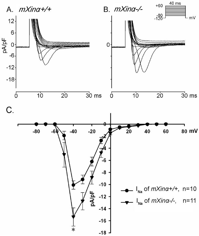
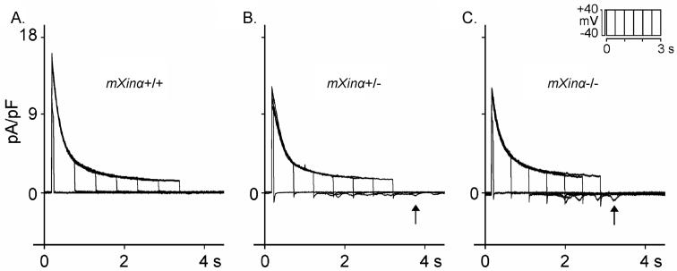
Methods: The present study aims to characterize changes in Na+ currents on depolarization and transient inward currents (Iti) on repolarization. Conduction velocity (CV) on the frontal surface of ventricles were also measured and compared.
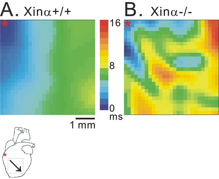
Results: Results of optical mapping on the Langendorff-perfused hearts at 37oC revealed a 36% reduction of CV in mXin alpha -/- ventricle. Pacing (3 Hz)-induced tachyarrhythmias were more frequently found and ventricular fibrillation (VF, 21 Hz for 5 min) occurred in one out of 8 mXin alpha-/- heart. When perfused at 30 degrees C, no VF was observed in both types of preparations. Voltage-clamp study on isolated ventricular myocytes at 37 degrees C shows increase in INa and Iti in mXin alpha -/- cardiomyocytes thus could explain the occurrence of re-entrant triggered arrhythmias.
Conclusion: The present results revealed that the CV was slower, but INa and Iti were increased in mXin alpha -/-cardiomyocytes thus were prone to reentrant triggered arrhythmias. Hypothermia could reduce the occurrence of arrhythmias.
Figures
References
-
- Wang DZ, Reiter RS, Lin JL, Wang Q, Williams HS, Krob SL, et al. Requirement of a novel gene, Xin, in cardiac morphogenesis. Development. 1999;126:1281–94. - PubMed
-
- Gustafson-Wagner EA, Lin JLC, Sinn H, Wang DZ, Reiter RS, Yang B, et al. Targeted deletion of mXinα gene, encoding an intercalated disc protein, leads to cardiac hypertrophy (Abstract) AHA Scientific Sessions 2006; 2006 Nov 12-15; Chicago, IL, USA; Circulation. 2006;114(Suppl):II–54.
-
- Cheng CP, Loh YX, Lin CI, Lai YJ, Chen YC, Sytwu HK, et al. In: Kimchi A, editor. Electrophysiological characteristics of ventricular myocytes of Xinα-deficient mice; Advances in Heart Disease. Proceedings of the 12th World Congress on Heart Disease; Vancouver, Canada; s.r.l. Bolongna, Italy; MEDIMOND. 2005 July 16-19.2005. pp. 25–9.
-
- Bers DM. Cardiac excitation-contraction coupling. Nature. 2002;415:198–205. - PubMed
-
- Wu SH, Chen YC, Higa S, Lin CI. Oscillatory transient inward currents in ventricular myocytes of healthy versus myopathic Syrian hamster. Clin Exp Pharmacol Physiol. 2004;31:668–76. - PubMed
Publication types
MeSH terms
Substances
Grants and funding
LinkOut - more resources
Full Text Sources
Medical
Miscellaneous