. 2007 Sep;148(9):4251-8.
doi: 10.1210/en.2006-0490. Epub 2007 Jun 21.

Side population cells in the mouse thyroid exhibit stem/progenitor cell-like characteristics

Affiliations

Side population cells in the mouse thyroid exhibit stem/progenitor cell-like characteristics

Nobuo Hoshi et al. Endocrinology. 2007 Sep.

Abstract

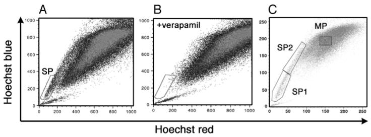
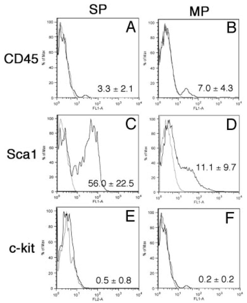
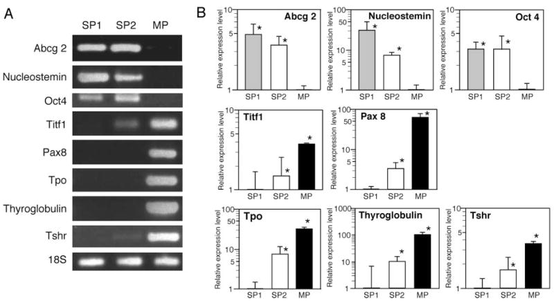
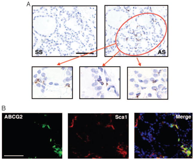
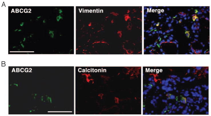
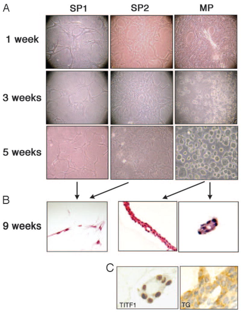
Side population (SP) cells are characterized by their ability to efflux the vital dye Hoechst 33342 (Sigma-Aldrich, St. Louis, MO) due to expression of the ATP binding cassette (ABC)-dependent transporter ABCG2, and are highly enriched for stem/progenitor cell activity. In this study we identified SP cells in murine thyroid, which are composed of two populations of cells: CD45(-)/c-kit(-)/Sca1(+) and CD45(-)/c-kit(-)/Sca1(-) cells. Quantitative RT-PCR analysis revealed that SP cells highly express ABCG2 and the stem cell marker genes encoding nucleostemin and Oct4, whereas the expression of genes encoding the thyroid differentiation markers, thyroid peroxidase, thyroglobulin (TG), and TSH receptor, and two transcription factors, thyroid transcription factor 1 (TITF1) and paired PAX8, critical for thyroid specific gene expression, are low in SP cells as compared with the main population cells. In situ hybridization and double immunofluorescence demonstrated that cells expressing Abcg2 gene reside in the interfollicular space of the thyroid gland. Approximately half and a small percentage of the ABCG2-positive cells were also positive for vimentin and calcitonin, respectively. After 9 wk under three-dimensional thyroid primary culture conditions, main population cells formed an epithelial arrangement and follicle-like structures that are immunoreactive for TITF1 and TG. In contrast, SP cells demonstrated very few morphological changes without any epithelial or follicle-like structure and negative immunostaining for TITF1 and TG. These results demonstrate that thyroid possesses SP cells that may represent stem/progenitor cells.


Figures

Fig. 1
Fig. 1
Detection of SP cells in adult mouse thyroid. Hoechst 33342 staining of mouse thyroid cell suspensions revealed the SP pattern of staining within the gated area (A), which was eliminated by treatment with verapamil (B). For some experiments, the SP fraction was further subdivided into SP1 and SP2 by setting a gate where the number of cells in the two regions becomes equal (C). Note that AB vs. C are two different thyroid preparations.
Fig. 2
Fig. 2
Expression of stem cell markers in thyroid SP and MP populations. Representative FACS results from three to eight independent experiments are shown for CD45 (A and B), Sca1 (C and D), and c-kit (E and F), and the mean number of percentage of (%) positive cells ± SD from these experiments are displayed at the lower right corner. MP cells are basically negative for all three markers, whereas approximately half the SP cells are positive for Sca1.
Fig. 3
Fig. 3
Gene expression patterns in thyroid SP and MP cells. RNAs isolated from SP1, SP2, and MP fraction of cells were subjected to RT-PCR, followed by agarose gel electrophoresis (representative results are shown in A) and quantitative RT-PCR analysis (B) for various genes as indicated. B, Expression levels are shown as a relative ratio in logarithmic scale based on the level of MP cells as one for Abcg2, nucleostemin and Oct 4, and the expression levels of SP1 cells as one for Titf1, Pax 8, Tpo, Tg, and TSHr. Note that the y-axis has different scales depending on the gene. Values are the mean ± SD from four different experiments, each performed in duplicate. *, P < 0.05. Note that in A, some images were intensified by Photoshop (Adobe Systems, Inc., San Jose, CA) to show clearly the presence of bands.
Fig. 4
Fig. 4
ABCG2 expression in adult mouse thyroid by in situ hybridization and immunofluorescence. A, In situ hybridization with sense (SS) and anti-sense Abcg2 probes (AS) was performed to locate the expression of Abcg2. Positive signals were detected only in cells surrounding thyroid follicles, but not cells lining follicles (lower panel). B, Immunofluorescence revealed that some cells are positive for only ABCG2 or Sca1, whereas others are double positive for both genes. Only representative areas are shown in which double-positive cells are present. A, upper panel: magnification, ×200; bar, 100 μm. A, lower panel, B: magnification, ×400; and bar, 50 μm.
Fig. 5
Fig. 5
Double-immunofluorescence study. Immunofluorescence was performed to detect double-positive cells for ABCG2 and vimentin (A), and ABCG2 and calcitonin (B). Only representative areas are shown in which double-positive cells are present. Magnification, ×400. Bar, 50 μm.
Fig. 6
Fig. 6
Morphological assessment of mouse thyroid SP and MP cells. SP1, SP2, and MP fractions were cultured using a three-dimensional collagen gel system, and cell morphology was recorded after 1, 3, and 5-wk culture (A). After 9 wk, cells were analyzed by hematoxylin and eosin staining for histology (B) and by immunohistochemistry (C) for TITF1 and TG. Only MP cells, but not SP1 or SP2 cells, developed epithelial arrangement and follicle-like structures that express TITF1 and TG. Magnification: ×100 (A), ×200 (B), and ×400 (C).

References

    1. Goodell MA, Brose K, Paradis G, Conner AS, Mulligan RC. Isolation and functional properties of murine hematopoietic stem cells that are replicating in vivo. J Exp Med. 1996;183:1797–1806. - PMC - PubMed
    1. Zhou S, Schuetz JD, Bunting KD, Colapietro AM, Sampath J, Morris JJ, Lagutina I, Grosveld GC, Osawa M, Nakauchi H, Sorrentino BP. The ABC transporter Bcrp1/ABCG2 is expressed in a wide variety of stem cells and is a molecular determinant of the side-population phenotype. Nat Med. 2001;7:1028–1034. - PubMed
    1. Asakura A, Rudnicki MA. Side population cells from diverse adult tissues are capable of in vitro hematopoietic differentiation. Exp Hematol. 2002;30:1339–1345. - PubMed
    1. Iwatani H, Ito T, Imai E, Matsuzaki Y, Suzuki A, Yamato M, Okabe M, Hori M. Hematopoietic and nonhematopoietic potentials of Hoechst(low)/side population cells isolated from adult rat kidney. Kidney Int. 2004;65:1604–1614. - PubMed
    1. Hussain SZ, Strom SC, Kirby MR, Burns S, Langemeijer S, Ueda T, Hsieh M, Tisdale JF. Side population cells derived from adult human liver generate hepatocyte-like cells in vitro. Dig Dis Sci. 2005;50:1755–1763. - PMC - PubMed

Publication types

MeSH terms