. 2007 Jul 3;104(27):11382-7.
doi: 10.1073/pnas.0704853104. Epub 2007 Jun 21.

T cell-specific deletion of the inositol phosphatase SHIP reveals its role in regulating Th1/Th2 and cytotoxic responses

Affiliations

T cell-specific deletion of the inositol phosphatase SHIP reveals its role in regulating Th1/Th2 and cytotoxic responses

Tatyana Tarasenko et al. Proc Natl Acad Sci U S A. .

Abstract

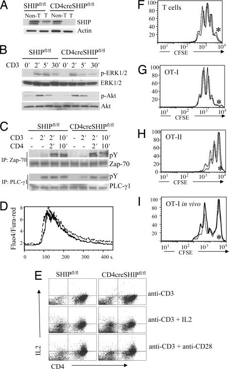
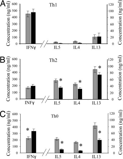
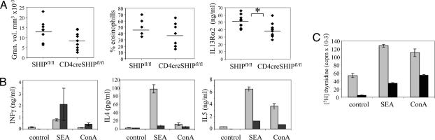
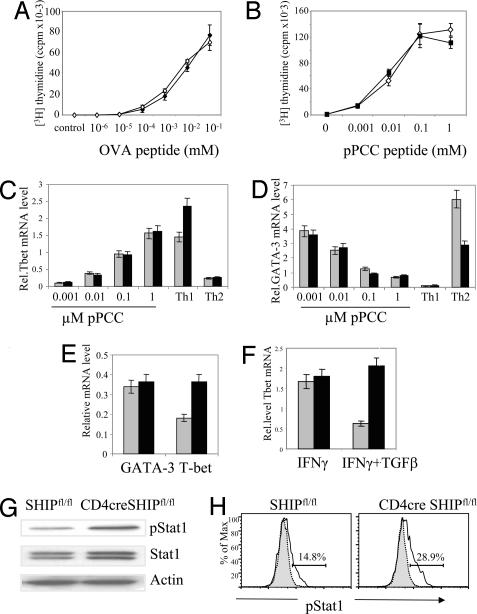
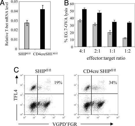
The 5'-phosphoinositol phosphatase SHIP negatively regulates signaling pathways triggered by antigen, cytokine and Fc receptors in both lymphocytes and myeloid cells. Mice with germ-line (null) deletion of SHIP develop a myeloproliferative-like syndrome that causes early lethality. Lymphocyte anomalies have been observed in SHIP-null mice, but it is unclear whether they are due to an intrinsic requirement of SHIP in these cells or a consequence of the severe myeloid pathology. To precisely address the function of SHIP in T cells, we have generated mice with T cell-specific deletion of SHIP. In the absence of SHIP, we found no differences in thymic selection or in the activation state and numbers of regulatory T cells in the periphery. In contrast, SHIP-deficient T cells do not skew efficiently to Th2 in vitro. Mice with T cell-specific deletion of SHIP show poor antibody responses on Alum/NP-CGG immunization and diminished Th2 cytokine production when challenged with Schistosoma mansoni eggs. The failure to skew to Th2 responses may be the consequence of increased basal levels of the Th1-associated transcriptional factor T-bet, resulting from enhanced sensitivity to cytokine-mediated T-bet induction. SHIP-deficient CD8(+) cells show enhanced cytotoxic responses, consistent with elevated T-bet levels in these cells. Overall our experiments indicate that in T cells SHIP negatively regulates cytokine-mediated activation in a way that allows effective Th2 responses and limits T cell cytotoxicity.


Conflict of interest statement

The authors declare no conflict of interest.

Figures

Fig. 1.
Fig. 1.
T cell-specific deletion of SHIP does not affect TCR signaling, in vitro T cell activation, or in vivo antigen-driven expansion. (A) Western blot with SHIP antibody shows efficient reduction in expression of SHIP protein in splenic T cells of CD4cre SHIPfl/fl mice. (B and C) No difference in Erk, Akt, Zap-70, or PLCγ1 activation on anti-CD3 or anti-CD3+ anti-CD4 stimulation of CD4cre SHIPfl/fl vs. SHIPfl/fl control T cells. (D) Equal calcium influx levels triggered by anti-CD3 stimulation of CD4cre SHIPfl/fl (solid line) vs. SHIPfl/fl T cells (dashed line). (E) CD4cre SHIPfl/fl T cells show the same level of IL2 production as SHIPfl/fl control cells. Purified naive CD4+ cells were incubated for 3 days with the indicated stimulus and tested for IL2 production by intracellular staining with FITC IL2 antibodies. (F–H) In vitro proliferation of CD4cre SHIPfl/fl (solid line) or SHIPfl/fl control (dashed line) T cells alone (F) or expressing the OT-I (G) and OT-II (H) TCR transgenes. CFSE-labeled cells were incubated on anti-CD3/CD28 plates for 3 days. Asterisks mark CFSE levels of undivided cells. (I) In vivo expansion of OT-I CD4cre SHIPfl/fl (solid line) or OT-I SHIPfl/fl control (dashed line) T cells. C57BL/6 host mice were injected with ovalbumin 1 day after receiving CFSE-labeled T cells from the indicated mice. Two days later, cells were purified from the draining lymph nodes, stained with CD8-Cychrome and Vα2-PE, and analyzed by FACS. Experiments were repeated three times, with three mice per group.
Fig. 2.
Fig. 2.
Humoral response in CD4cre SHIPfl/fl (filled triangles) and SHIPfl/fl (open squares) mice. (A) Mice were immunized with 50 μg of NP-CGG with Alum and pertussis toxin, and the levels of NP-specific antibodies of different subclasses in the serum were determined 2 weeks after the primary immunization (Left) or 1 week after reimmunization given on day 28 (Right). Serum was diluted 1:5,000 for primary immunizations and 1:100,000 for secondary immunizations. Lines indicate the mean antibody response. (B) Mice were immunized with 50 μg of TNP-KLH with RIBI adjuvant, and TNP-specific antibody levels in the serum were determined as in A. (C) Impaired germinal center formation in CD4cre SHIPfl/fl (Right) vs. control mice (Left) after immunization with NP-CGG plus Alum plus pertussis toxin. Gated for B220-positive cells. Results are representative from three independent experiments. (D) Cytokine production by cells purified from NP-CGG/Alum-challenged CD4cre SHIPfl/fl (black) and SHIPfl/fl mice (gray). Lymph node cells were isolated on day 7 after reimmunization and stimulated with NP-BSA for 24 h.
Fig. 3.
Fig. 3.
SHIP-deficient CD4 T cells polarize in culture but have decreased Th2 cytokine production. CD4cre SHIPfl/fl (dark bars) and SHIPfl/fl (light bars) CD4+ T cells were stimulated with anti-CD3 plus anti-CD28 under Th1 (A), Th2 (B), or Th0 (C) polarizing conditions for 3 days, expanded in IL2 for 4 days, and restimulated with anti-CD3 plus anti-CD28. Supernatants were collected 24 h after restimulation and cytokine expression assessed by the CBA Assay. ∗, P < 0.05.
Fig. 4.
Fig. 4.
Altered responses to the Th2 polarizing agent S. mansoni eggs in mice with T cell-specific deletion of SHIP. (A) Lung granuloma volume, percentage granulome eosinophils, and serum IL13Rα2 from CD4cre SHIPfl/fl and SHIPfl/fl mice challenged with S. mansoni eggs. ∗, P < 0.05. (B) Reduced Th2 cytokine production by T cells purified from infected CD4cre SHIPfl/fl mice (dark bars) vs. SHIPfl/fl controls (light bars). Draining mediastinal lymph nodes were dissected 10 days after secondary injection with S. mansoni eggs. Cells from five mice were pooled and cultured with ConA or SEA antigen for 72 h, and supernatants were analyzed for cytokine production. (C) Reduced SEA or ConA-induced proliferation of T cells purified from lymph nodes of infected CD4cre SHIPfl/fl mice (dark bars) vs. SHIPfl/fl controls (light gray bars).
Fig. 5.
Fig. 5.
Increased basal level of T-bet in SHIP-deleted T cells due to altered cytokine signaling. (A) Proliferation of naive OT-II CD4cre SHIPfl/fl T cells (filled diamonds) vs. OT-II SHIPfl/fl controls (empty diamonds) in the presence of irradiated splenocytes and the indicated concentration of OVA peptide. (B) Proliferation of naive 5CC7 CD4cre SHIPfl/fl T cells (filled squares) vs. 5CC7 SHIPfl/fl controls (empty triangles) in the presence of irradiated P13.9 fibroblasts as described in Methods. (C and D) SHIP deletion does not alter T-bet or GATA-3 induction on antigen stimulation of T cells. Naive CD4+ CD62L+ cells from 5CC7 CD4cre SHIPfl/fl mice (black bars) and 5CC7 SHIPfl/fl control mice (gray bars) were stimulated with mitomycin C-treated P13.9 fibroblast cells that had been preloaded with 0.001–1 μM pPCC. Cells were harvested after 24 h of incubation, and relative mRNA levels (compared with L32 gene) for T-bet (C) and GATA-3 (D) were measured by real-time PCR. The experiment was performed twice with the same result. (E) T-bet and GATA-3 mRNA expression in naive freshly isolated CD4+ CD62L+ T cells from CD4cre SHIPfl/fl (black bars) and SHIPfl/fl mice (gray bars). (F) SHIP mediates TGFβ1 inhibition of IFNγ-induced T-bet up-regulation. Purified CD4+ CD62L+ T cells were incubated with IFNγ with or without TGFβ for 24 h, and the relative level of mRNA T-bet was assessed by real-time PCR. (G) Increased STAT phosphorylation in freshly purified T cells from CD4cre SHIPfl/fl vs. control SHIPfl/fl mice. (H) Increased STAT phosphorylation in IFNγ-activated T cells from CD4cre SHIPfl/fl (Right) vs. control SHIPfl/fl mice (Left). Th1-skewed T cells were incubated for 24 h with IL12 to up-regulate the IFNγ receptor level, rested for 4 h, and incubated with IFNγ for 15 min. The proportion of cells expressing pSTAT1 was calculated as the difference between anti-pSTAT intracellular flow cytometry (solid line) minus unstimulated control (gray blot).
Fig. 6.
Fig. 6.
Enhanced CTL function of SHIP-deleted CD8+ cells. (A) SHIP-deficient CD8+ T cells express 60% more T-bet mRNA. Real-time PCR was performed on mRNA from purified splenic CD8+ cells. Values are relative to the expression of L32 gene. (B) CD4cre SHIPfl/fl and SHIPfl/fl OT-I cells were stimulated with irradiated splenocytes plus peptide for 72 h, harvested, rested, incubated for 6 h with a mixture of EL-4 cells, labeled with high levels of CFSE and EG7-OVA cells, and labeled with low levels of CFSE. The ratio of the numbers of two populations of cells were determined by flow cytometry at the end of the incubation period. The percentage of killing was calculated as 1 minus the ratio of target to control over the ratio of target to control in the absence of effector T cells. (C) Granzyme-dependent killing of EG-7 OVA targets by CD4cre SHIPfl/fl (Right) and SHIPfl/fl (Left) OT-I cells measured by using the GranToxiLux kit. EG7-OVA cells are FL-4+. Upper left quadrants represent viable target cells, whereas upper right quadrants represent dying, VGPD'FGR substrate-positive target cells. Effector cells occupy the lower two quadrants.

References

    1. Ono M, Okada H, Bolland S, Yanagi S, Kurosaki T, Ravetch JV. Cell. 1997;90:293–301. - PubMed
    1. Kalesnikoff J, Sly LM, Hughes MR, Buchse T, Rauh MJ, Cao LP, Lam V, Mui A, Huber M, Krystal G. Rev Physiol Biochem Pharmacol. 2003;149:87–103. - PubMed
    1. Rauh MJ, Kalesnikoff J, Hughes M, Sly L, Lam V, Krystal G. Biochem Soc Trans. 2003;31:286–291. - PubMed
    1. Rohrschneider LR, Fuller JF, Wolf I, Liu Y, Lucas DM. Genes Dev. 2000;14:505–520. - PubMed
    1. Ono M, Bolland S, Tempst P, Ravetch JV. Nature. 1996;383:263–266. - PubMed

Publication types

MeSH terms

Substances