Review
. 2007 Oct;293(4):H2024-38.
doi: 10.1152/ajpheart.00355.2007. Epub 2007 Jun 22.

Role of spatial dispersion of repolarization in inherited and acquired sudden cardiac death syndromes

Affiliations
Review

Role of spatial dispersion of repolarization in inherited and acquired sudden cardiac death syndromes

Charles Antzelevitch. Am J Physiol Heart Circ Physiol. 2007 Oct.

Abstract

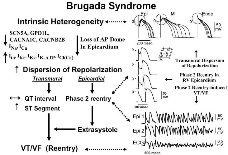
This review examines the role of spatial electrical heterogeneity within the ventricular myocardium on the function of the heart in health and disease. The cellular basis for transmural dispersion of repolarization (TDR) is reviewed, and the hypothesis that amplification of spatial dispersion of repolarization underlies the development of life-threatening ventricular arrhythmias associated with inherited ion channelopathies is evaluated. The role of TDR in long QT, short QT, and Brugada syndromes, as well as catecholaminergic polymorphic ventricular tachycardia (VT), is critically examined. In long QT syndrome, amplification of TDR is often secondary to preferential prolongation of the action potential duration (APD) of M cells; in Brugada syndrome, however, it is thought to be due to selective abbreviation of the APD of the right ventricular epicardium. Preferential abbreviation of APD of the endocardium or epicardium appears to be responsible for the amplification of TDR in short QT syndrome. In catecholaminergic polymorphic VT, reversal of the direction of activation of the ventricular wall is responsible for the increase in TDR. In conclusion, long QT, short QT, Brugada, and catecholaminergic polymorphic VT syndromes are pathologies with very different phenotypes and etiologies, but they share a common final pathway in causing sudden cardiac death.


Figures

Figure 1
Figure 1
Ionic distinctions among epicardial, M and endocardial cells. A: Action potentials recorded from myocytes isolated from the epicardial, endocardial and M regions of the canine left ventricle. B: I-V relations for IK1 in epicardial, endocardial and M region myocytes. Values are mean ± S.D. C: Transient outward current (Ito) recorded from the three cell types. D: The average peak current-voltage relationship for Ito for each of the three cell types. Values are mean±S.D. E: Voltage-dependent activation of the slowly activating component of the delayed rectifier K+ current (IKs) (currents were elicited by the voltage pulse protocol shown in the inset; Na+-, K+- and Ca2+- free solution). F: Voltage dependence of IKs (current remaining after exposure to E-4031) and IKr (E-4031-sensitive current). Values are mean ± S.E. * p<0.05 compared with Epi or Endo. From references (61; 62; 139) with permission. G: Reverse-mode sodium-calcium exchange currents recorded in potassium- and chloride-free solutions at a voltage of −80 mV. INa-Ca was maximally activated by switching to sodium-free external solution at the time indicated by the arrow. H: Midmyocardial sodium-calcium exchanger density is 30% greater than endocardial density, calculated as the peak outward INa-Ca normalized by cell capacitance. Endocardial and epicardial densities were not significantly different. I: TTX-sensitive late sodium current. Cells were held at −80 mV and briefly pulsed to −45 mV to inactivate fast sodium current before stepping to −10 mV. J: Normalized late sodium current measured 300 msec into the test pulse was plotted as a function of test pulse potential. Modified from reference (139) with permission.
Figure 2
Figure 2
Transmembrane activity recorded from cells isolated from the epicardial (Epi), M and endocardial (Endo) regions of the canine left ventricle at basic cycle lengths (BCL) of 300 to 5000 msec (steady-state conditions). The M and transitional cells were enzymatically dissociated from the midmyocardial region. Deceleration- induced prolongation of APD in M cells is much greater than in epicardial and endocardial cells. The spike and dome morphology is also more accentuated in the epicardial cell. From (62), with permission.
Figure 3
Figure 3
Transmural distribution of action potential duration and tissue resistivity across the ventricular wall. A: Schematic diagram of the coronary-perfused canine LV wedge preparation. Transmembrane action potentials are recorded simultaneously from epicardial (Epi), M region (M) and endocardial (Endo) sites using three floating microelectrodes. A transmural ECG is recorded along the same transmural axis across the bath, registering the entire field of the wedge. B: Histology of a transmural slice of the left ventricular wall near the epicardial border. The region of sharp transition of cell orientation coincides with the region of high tissue resistivity depicted in panel D and the region of sharp transition of action potential duration illustrated in panel C. C: Distribution of conduction time (CT), APD90 and repolarization time (RT = APD90 + CT) in a canine left ventricular wall wedge preparation paced at BCL of 2000 msec. A sharp transition of APD90 is present between epicardium and subepicardium. Epi: epicardium; M: M Cell; Endo: endocardium. RT: repolarization time; CT: conduction time. D: Distribution of total tissue resistivity (Rt) across the canine left ventricular wall. Transmural distances at 0% and 100% represent epicardium and endocardium, respectively. * p<0.01 compared with Rt at mid-wall. Tissue resistivity increases most dramatically between deep subepicardium and epicardium. Error bars represent SEM (n=5). From (11; 134) with permission.
Figure 4
Figure 4
LQT1, LQT2, and LQT3 models of LQTS. Panels A–C shows action potentials simultaneously recorded from endocardial (Endo), M and epicardial (Epi) sites of arterially-perfused canine left ventricular wedge preparations together with a transmural ECG. BCL = 2000 msec. Transmural dispersion of repolarization across the ventricular wall, defined as the difference in the repolarization time between M and epicardial cells, is denoted below the ECG traces. LQT1 model was mimicked using Isoproterenol + chromanol 293B − an IKs blocker. LQT2 was created using the IKr blocker d-sotalol + low [K+]o. LQT3 was mimicked using the seas anemone toxin ATX-II to augments late INa. Panels D–F: Effect of isoproterenol in the LQT1, LQT2 and LQT3 models. In LQT1, isoproterenol (Iso) produces a persistent prolongation of the APD90 of the M cell and of the QT interval (at both 2 and 10 minute), whereas the APD90 of the epicardial cell is always abbreviated, resulting in a persistent increase in TDR (D). In LQT2, isoproterenol initially prolongs (2 minute) and then abbreviates the QT interval and the APD90 of the M cell to the control level (10 minute), whereas the APD90 of epicardial cell is always abbreviated, resulting in a transient increase in TDR (E). In LQT3, isoproterenol produced a persistent abbreviation of the QT interval and the APD90 of both M and epicardial cells (at both 2 and 10 minute), resulting in a persistent decrease in TDR (F). *P < .0005 vs. Control; † P < .0005, †† P < .005, ††† P < .05, vs. 293B, d-Sotalol (d-Sot) or ATX-II. (Modified from references (86; 87; 90)with permission).
Figure 5
Figure 5
Proposed cellular mechanism for the development of Torsade de Pointes in the long QT syndromes.
Figure 6
Figure 6
Cellular basis for electrocardiographic and arrhythmic manifestation of Brugada Syndrome. Each panel shows transmembrane action potentials from one endocardial (top) and two epicardial sites together with a transmural ECG recorded from a canine coronary-perfused right ventricular wedge preparation. A: Control (BCL 400 msec). B: Combined sodium and calcium channel block with terfenadine (5 μM) accentuates the epicardial action potential notch creating a transmural voltage gradient that manifests as a ST segment elevation or exaggerated J wave in the ECG. C: Continued exposure to terfenadine results in all-or-none repolarization at the end of phase 1 at some epicardial sites but not others, creating a local epicardial dispersion of repolarization (EDR) as well as a transmural dispersion of repolarization (TDR). D: Phase 2 reentry occurs when the epicardial action potential dome propagates from a site where it is maintained to regions where it has been lost giving rise to a closely coupled extrasystole. E: Extrastimulus (S1-S2 = 250 msec) applied to epicardium triggers a polymorphic VT. F: Phase 2 reentrant extrasystole triggers a brief episode of polymorphic VT. (Modified from reference (48), with permission)
Figure 7
Figure 7
Proposed mechanism for the Brugada syndrome. A shift in the balance of currents serves to amplify existing heterogeneities by causing loss of the action potential dome at some epicardial, but not endocardial sites. A vulnerable window develops as a result of the dispersion of repolarization and refractoriness within epicardium as well as across the wall. Epicardial dispersion leads to the development of phase 2 reentry, which provides the extrasystole that captures the vulnerable window and initiates VT/VF via a circus movement reentry mechanism. Modified from (4), with permission.
Figure 8
Figure 8
Proposed mechanism for arrhythmogenesis in the short QT syndrome. An increase in net outward current due to a reduction in late inward current or augmentation of outward repolarizing current serves to abbreviate action potential duration heterogeneously leading to an amplification of transmural dispersion of repolarization and the creation of a vulnerable window for the development of reentry. Reentry is facilitated both by the increase in TDR and abbreviation of refractoriness.
Figure 9
Figure 9
The role of transmural dispersion of repolarization (TDR) in channelopathy-induced sudden cardiac death. In the long QT syndrome, QT increases as a function of disease or drug concentration. In the Brugada syndrome it remains largely unchanged and in the short QT syndrome QT interval decreases as a function of disease or drug. The three syndromes have in common the ability to amplify TDR, which results in the development of TdP when dispersion reaches the threshold for reentry. The threshold for reentry decreases as APD and refractoriness are reduced. Modified from (12), with permission.

References

    1. Aiba T, Shimizu W, Hidaka I, Uemura K, Noda T, Zheng C, Kamiya A, Inagaki M, Sugimachi M, Sunagawa K. Cellular basis for trigger and maintenance of ventricular fibrillation in the Brugada syndrome model: high-resolution optical mapping study. J Am Coll Cardiol. 2006;47:2074–2085. - PubMed
    1. Akar FG, Rosenbaum DS. Transmural electrophysiological heterogeneities underlying arrhythmogenesis in heart failure. Circ Res. 2003;93:638–645. - PubMed
    1. Akar FG, Tomaselli GF. Conduction abnormalities in nonischemic dilated cardiomyopathy: basic mechanisms and arrhythmic consequences. Trends Cardiovasc Med. 2005;15:259–264. - PubMed
    1. Antzelevitch C. The Brugada Syndrome: Diagnostic Criteria and Cellular Mechanisms. Eur Heart J. 2001;22:356–363. - PubMed
    1. Antzelevitch C. The Brugada syndrome: ionic basis and arrhythmia mechanisms. J Cardiovasc Electrophysiol. 2001;12:268–272. - PubMed

Publication types

MeSH terms

LinkOut - more resources