. 2007 Aug;6(8):1310-9.
doi: 10.1128/EC.00136-07. Epub 2007 Jun 22.

Development of a gene knockout system in Candida parapsilosis reveals a conserved role for BCR1 in biofilm formation

Affiliations

Development of a gene knockout system in Candida parapsilosis reveals a conserved role for BCR1 in biofilm formation

Chen Ding et al. Eukaryot Cell. 2007 Aug.

Abstract

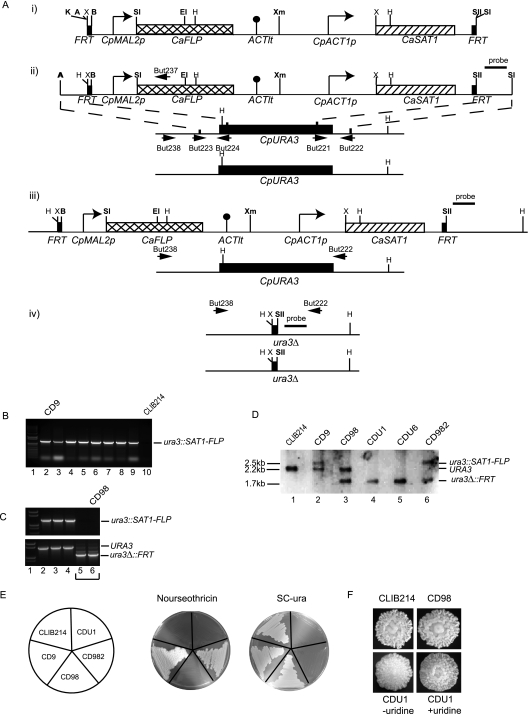
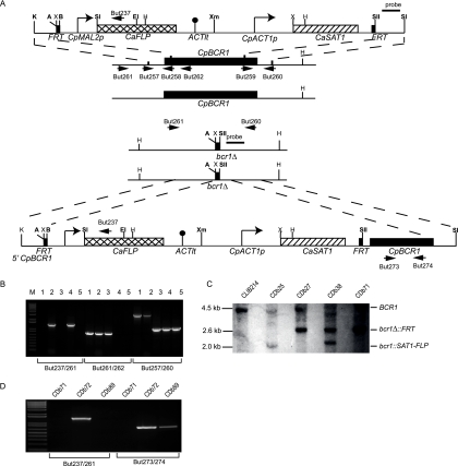
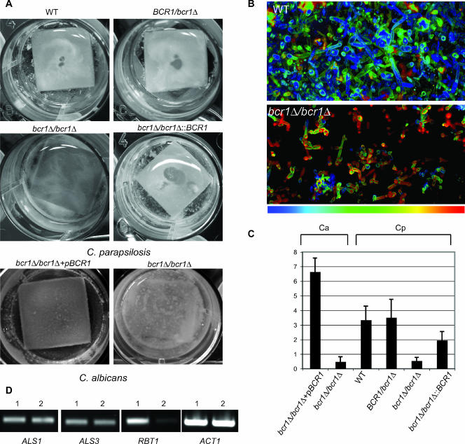
Candida parapsilosis is an important cause of candidiasis, yet few molecular tools are available. We adapted a recyclable nourseothricin resistance marker gene (SAT1) originally developed for use with C. albicans in order to generate gene knockouts from C. parapsilosis. We first replaced the promoters driving expression of the FLP recombinase and the SAT1 genes with the equivalent sequences from C. parapsilosis. We then used the cassette to generate a homozygous knockout of C. parapsilosis URA3. The ura3 knockouts have altered colony morphologies. We also knocked out both alleles of an ortholog of BCR1, a gene encoding a transcription factor known to be required for biofilm development in C. albicans. We show that C. parapsilosis BCR1 is necessary for biofilm development in C. parapsilosis and for expression of the cell wall protein encoded by RBT1. Our results suggest that there are significant similarities in the regulation of biofilms between the two species, despite the fact that C. parapsilosis does not generate true hyphae and that BCR1 regulates the expression of many hypha-specific adhesins in C. albicans.


Figures

FIG. 1.
FIG. 1.
Construction of a Cpura3Δ strain. (A) (i) A disruption cassette was adapted from the SAT1 flipper system described by Reuss et al. (43) by replacing the MAL2 and ACT1 promoters with equivalent sequences from C. parapsilosis. (ii) Upstream and downstream sequences from CpURA3 were amplified using primer pairs But223/But224 and But221/But222 and inserted at the ApaI/XhoI and SacII/SacI sites surrounding the flipper cassette. (iii) The entire cassette and flanking regions were excised by digestion with ApaI and SacI and targeted to the CpURA3 alleles by homologous recombination. The figure shows the structure of the cassette integrated at one allele. (iv) The cassette was recycled from the first allele by induction of the FLP recombinase, inserted at the second allele and recycled once more. Restriction sites shown in bold are unique. A, ApaI; B, BamHI; E, EcoRI; H, HindIII; K, KpnI; SI, SacI; SII, SacII; Sl, SalI; X, XhoI; Xm, XmaI. (B) Insertion of the SAT1 cassette at the first CpURA3 allele was verified by PCR using primers But237 (from within the FLP gene) and But238 (from upstream of CpURA3, outside the range of the deletion). Lanes 2 to 9 show the presence of a 1.3-kb fragment in eight independent disruptants that is not present in the wild-type strain CLIB214 (lane 10). One (lane 2, strain CD9) was chosen to recycle the cassette. Lane 1 contains a molecular size marker (1 kb plus; Invitrogen). (C) Recycling of the SAT1-FLP cassette. PCR amplification using primers But237 and But238 shows that the strains in lanes 2, 3, and 4 have not lost the cassette, whereas those in lanes 5 and 6 have (top panel). This is confirmed in the bottom panel, which shows PCR amplification using the primer pair But238 and But222 surrounding CpURA3. The strains in lanes 2, 3, and 4 contain the wild-type CpURA3 allele (1.5 kb), whereas those in lanes 5 and 6 have the deleted allele (1.0 kb). One isolate (CD98) was chosen for disruption of the second allele. (D) The structure of the disruption was also confirmed by Southern hybridization using the probe indicated, derived from the downstream region of CpURA3. Genomic DNA was digested with HindIII. The probe hybridizes to one fragment of 2.2 kb (URA3) in the starting strain CLIB214 (lane 1), representing the wild-type allele. Introducing the cassette at one allele produces one larger band of 2.5 kb (ura3::SAT1-FLP) and one wild-type band (CD9, lane 2). Recycling the cassette results in a small fragment of 1.7 kb (ura3Δ::FRT), corresponding to the deleted allele, and a wild-type fragment (CD98, lane 3). The cassette was introduced at the second allele, generating both large (ura3::SAT1-FLP) and small (ura3Δ::FRT) fragments (CD982, lane 6). Recycling the cassette resulted in only one fragment, corresponding to the deleted CpURA3 allele (ura3Δ::FRT; CDU1 and CDU6, lanes 4 and 5). (E) The strains containing SAT1-FLP integrated at the first allele of CpURA3 (CD9) or the second allele (CD982) are resistant to nourseothricin (at 200 μg ml−1). All other strains are sensitive. The strains in which both alleles of CpURA3 were deleted (CDU1 and CDU6) failed to grow in the absence of uracil. (F) The phenotypes of the wild-type (CLIB214) and the heterozygote knockout strains (CD98) are the same when grown on YPD medium and resemble the crater phenotype described previously (26). The double-deletion strain (CDU1) has a different appearance, which reverts to the wild-type phenotype when excess uridine (50 μg ml−1) is added.
FIG. 2.
FIG. 2.
Construction of a Cpbcr1Δ strain. (A) Upstream and downstream sequences from CpBCR1 were amplified using primer pairs But257/But258 and But259/But260 and inserted at the KpnI/ApaI and SacII/SacI sites surrounding the SAT flipper cassette. The entire cassette and flanking regions were excised by digestion with KpnI and SacI and targeted to the CpBCR1 alleles by homologous recombination. The cassette was recycled from the first allele by induction of the FLP recombinase, inserted at the second allele, and recycled once more. Restriction sites shown in bold are unique. A, ApaI; B, BamHI; E, EcoRI; H, HindIII; K, KpnI; SI, SacI; SII, SacII; Sl, SalI; X, XhoI; Xm, XmaI. The CpBCR1 gene was reintroduced at one deletion allele by inserting a 550-bp fragment from the CpBCR1 promoter (plus 45 codons) upstream of the SAT1-FLP cassette and the entire open reading frame (including 82 bp of the promoter) downstream of the cassette. The cassette was removed by recombination between the FRT sites, as described before. (B) Construction of the deletion was confirmed by PCR using three different primer combinations. The order of the strains for each reaction is as follows: lane 1, CLIB214; lane 2, CDb25 (first integration); lane 3, CDb27 (first recycle); lane 3, CDb38 (second integration); lane 5, CDb71 (second recycle). Amplification with primers But261/But237 generates a 1.5-kb fragment from within the FLP gene to upstream of BCR1. Primers But261/But262 amplify a 0.8-kb fragment from the 5′ region of the BCR1 gene. Primers But257/But260 amplify a 3.0-kb fragment from the wild-type BCR1 allele and a 1-kb fragment from the bcr1Δ allele. (C) The structure of the disruption was also confirmed by Southern hybridization using a probe derived from the downstream region of CpBCR1. Genomic DNA was digested with HindIII. The probe hybridizes to one 4.5-kb fragment (BCR1) in the starting strain, CLIB214 (lane 1), representing the wild-type allele. Introducing the cassette at one allele produces one smaller band of 2.0 kb (bcr1::SAT1-FLP) and one wild-type band (CDb25, lane 2). Recycling the cassette results in a fragment of 2.6 kb (bcr1Δ::FRT), corresponding to the deleted allele, and a wild-type fragment (CDb27, lane 3). The cassette was introduced at the second allele, generating both 2.0-kb (bcr1::SAT1-FLP) and 2.6-kb (bcr1Δ::FRT) fragments (CDb38, lane 4). Recycling the cassette results in only one fragment, corresponding to the deleted BCR1 allele (bcr1Δ::FRT; CDb71, lane 5). (D) Reintegration of the CpBCR1 gene at one deletion allele was confirmed by PCR. Amplification with primers But261/But237 generates a 1.5-kb fragment from within the FLP gene to upstream of BCR1 in CDb72 (containing the SAT1-FLP cassette and the entire CpBCR1 gene), with no amplification from the double-deletion (CDb71) or from the reconstituted strain following recycling of the SAT1 cassette. Primers But261/But237 amplify a 0.8-kb fragment from within the CpBCR1 gene before and after recycling the cassette (CDb72 and CDb89) but not in the deletion strain (CDb71).
FIG. 3.
FIG. 3.
Disrupting bcr1 impairs biofilm development. (A) C. parapsilosis wild-type (CLIB214), BCR1/bcr1Δ heterozygote (CDb27), bcr1Δ/bcr1Δ homozygote (CDb71), and reconstituted BCR1 (CDb89) strains were grown for 48 h in SD medium on silicone squares placed at the bottom of 12-well plates. Adherence to the silicone was monitored by photography and compared to that of the biofilms generated by C. albicans BCR1 reconstituted (CJN698) and bcr1/bcr1 disruption (CJN702) strains grown in Spider medium. Disrupting bcr1 greatly reduces biofilm formation in both species. (B) CLSM depth views. Biofilms were stained with conA and visualized by using a Zeiss LSM510 confocal scanning microscope, and false-color depths were constructed. Blue cells are closest to the silicone, and red cells are farthest away. The wild-type (C. parapsilosis CLIB214) biofilms are approximately 100 μm deep, and the bcr1Δ (C. parapsilosis CDb71) biofilms are approximately 20 μm deep. (C) The biomass of the biofilms generated was determined by measuring the dry weight. Averages of six biological replicates are shown. Ca, C. albicans; Cp, C. parapsilosis. Phenotypes are represented by strains as follows: bcr1Δ/bcr1Δ+pBCR1, C. albicans CJN698; bcr1Δ/bcr1Δ, C. albicans CJN702; wild type (WT), C. parapsilosis CLIB214; BCR1/bcr1Δ, C. parapsilosis CDb27; bcr1Δ/bcr1Δ, C. parapsilosis CDb71; bcr1Δ/bcr1Δ::BCR1, C. parapsilosis CDb89. The differences between the biofilms from wild-type and bcr1 knockouts (P < 0.0001) and reconstituted and bcr1 knockouts (P = 0.003) in C. parapsilosis are statistically significant (t test, two-tailed P value). (D) Expression levels of ALS1, ALS3, and RBT1 were compared in the C. parapsilosis wild-type (1, CLIB214) and C. parapsilosis bcr1Δ (2, CDb71) strains using RT-PCR. Actin levels (ACT1) were measured as a control.

Similar articles

Cited by

References

    1. Almirante, B., D. Rodriguez, M. Cuenca-Estrella, M. Almela, F. Sanchez, J. Ayats, C. Alonso-Tarres, J. L. Rodriguez-Tudela, and A. Pahissa. 2006. Epidemiology, risk factors, and prognosis of Candida parapsilosis bloodstream infections: case-control population-based surveillance study of patients in Barcelona, Spain, from 2002 to 2003. J. Clin. Microbiol. 44:1681-1685. - PMC - PubMed
    1. Bain, J. M., C. Stubberfield, and N. A. Gow. 2001. Ura-status-dependent adhesion of Candida albicans mutants. FEMS Microbiol. Lett. 204:323-328. - PubMed
    1. Bakir, M., N. Cerikcioglu, R. Barton, and A. Yagci. 2006. Epidemiology of candidemia in a Turkish tertiary care hospital. APMIS 114:601-610. - PubMed
    1. Barchiesi, F., G. Caggiano, L. Falconi Di Francesco, M. T. Montagna, S. Barbuti, and G. Scalise. 2004. Outbreak of fungemia due to Candida parapsilosis in a pediatric oncology unit. Diagn. Microbiol. Infect. Dis. 49:269-271. - PubMed
    1. Blankenship, J. R., and A. P. Mitchell. 2006. How to build a biofilm: a fungal perspective. Curr. Opin. Microbiol. 9:588-594. - PubMed

Publication types

LinkOut - more resources