. 2007;35(14):4597-607.
doi: 10.1093/nar/gkm468. Epub 2007 Jun 22.

Novel activity of eukaryotic translocase, eEF2: dissociation of the 80S ribosome into subunits with ATP but not with GTP

Affiliations

Novel activity of eukaryotic translocase, eEF2: dissociation of the 80S ribosome into subunits with ATP but not with GTP

Natalia Demeshkina et al. Nucleic Acids Res. 2007.

Erratum in

  • Nucleic Acids Res. 2007;35(20):7040

Abstract

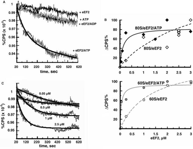
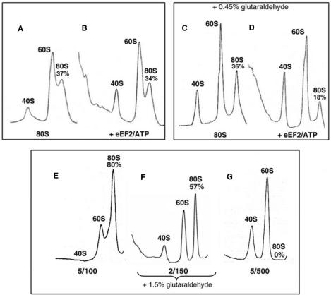
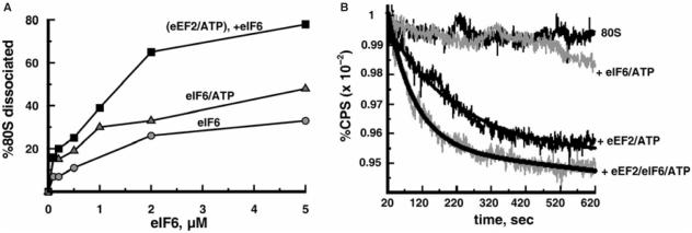
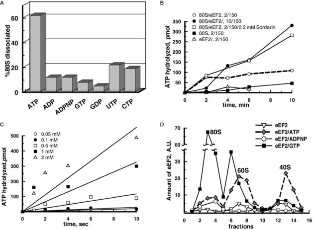
Ribosomes must dissociate into subunits in order to begin protein biosynthesis. The enzymes that catalyze this fundamental process in eukaryotes remained unknown. Here, we demonstrate that eukaryotic translocase, eEF2, which catalyzes peptide elongation in the presence of GTP, dissociates yeast 80S ribosomes into subunits in the presence of ATP but not GTP or other nucleoside triphosphates. Dissociation was detected by light scattering or ultracentrifugation after the split subunits were stabilized. ATP was hydrolyzed during the eEF2-dependent dissociation, while a non-hydrolyzable analog of ATP was inactive in ribosome splitting by eEF2. GTP inhibited not only ATP hydrolysis but also dissociation. Sordarin, a fungal eEF2 inhibitor, averted the splitting but stimulated ATP hydrolysis. Another elongation inhibitor, cycloheximide, also prevented eEF2/ATP-dependent splitting, while the inhibitory effect of fusidic acid on the splitting was nominal. Upon dissociation of the 80S ribosome, eEF2 was found on the subunits. We propose that the dissociation activity of eEF2/ATP plays a role in mobilizing 80S ribosomes for protein synthesis during the shift up of physiological conditions.


Figures

Figure 1.
Figure 1.
eEF2 dissociates 80S ribosomes in the presence of ATP: light scattering analysis. (A) Ribosomes (0.05 μM) were incubated with 2.5 μM eEF2 and 0.5 mM ATP in buffer 2/150 containing 0.8% glycerol. In the control experiments, the ribosomes were incubated with ATP or eEF2 alone or with eEF2/ADP. The light scattering (CPS) is expressed as the percentage of the initial value at 20 s after mixing and is plotted against the time of incubation. (B) The instantaneous increase in light scattering (ΔCPS) due to the binding of eEF2 to ribosomes was measured at 20 s after mixing of the ribosomes and eEF2 (or eEF2/ATP). ΔCPS is expressed as the percentage of the maximum light scattering increase observed at the point of ribosome saturation with eEF2, and is plotted against the amount of eEF2 added. Upper panel: binding of eEF2 to 80S ribosomes with (closed diamonds) and without (open diamond) ATP. Lower panel: binding of eEF2 to 60S subunits with (gray circles) and without (open circles) ATP. Experimental conditions were the same as in (A). (C) As in (A) except that various amounts of eEF2 (in the absence of glycerol) were added as indicated.
Figure 2.
Figure 2.
Splitting of 80S ribosomes by eEF2/ATP: sedimentation studies. (A and B) Ribosomes (0.05 μM) were incubated in buffer 2/150 alone (A) or with 1 µM eEF2 and 0.5 mM ATP (B) for 20 min at 30°C and subjected to SDGC. (C and D) Conditions in (C) and (D) were identical to (A) and (B), respectively, except that the reaction mixtures were treated with 0.45% glutaraldehyde (v/v) before SDGC. (E–G) The stock solution of ribosomes (0.5 μM in buffer 5/100) was diluted to a final concentration 0.05 μM in the buffers 5/100, 2/150 and 5/500. In buffer 2/150, ribosomes were treated with 1.5% glutaraldehyde (v/v). The ribosomes were then sedimented as in (A–D). The percentages of 80S ribosomes relative to the total ribosome content are indicated.
Figure 3.
Figure 3.
The 80S ribosomes transiently dissociated by eEF2/ATP are stabilized by eIF6. (A) Ribosomes (0.05 μM) were pre-incubated with 2.5 μM eEF2 and 0.5 mM ATP for 6 min at 30°C in buffer 2/150, then various amounts of eIF6 were added as indicated and incubated further for 15 min. The reaction mixtures were analyzed by SDGC. In the control experiments, ribosomes were exposed for 15 min to eIF6 or eIF6/ATP. Percentages of 80S ribosomes dissociated (Z) were calculated as follows: Z = 100 × (1 − (Y/W)), where (Y) is the amount of 80S ribosomes expressed as a percentage of total ribosomes remaining after the addition of factors and (W) is the amount of 80S ribosomes (percentage of total ribosomes) without the addition of factors. (B) Ribosomes (0.05 µM) were mixed with factors as indicated in buffer 2/150 containing 0.8% glycerol and the light scattering change was measured as in Figure 1A. Final concentrations of eEF2, eIF6 and ATP were 2.5 μM, 2.5 μM and 0.5 mM, respectively.
Figure 4.
Figure 4.
ATP but not other nucleotides are essential for the eEF2-dependent dissociation of 80S ribosomes and the binding of eEF2 to subunits: ATP hydrolysis during the splitting. (A) Ribosomes (0.05 μM) were pre-incubated in buffer 2/150 with 2.5 μM eEF2 and 0.5 mM of one of the specified nucleotides for 6 min at 30°C and then eIF6 was added to 2 μM and further incubated for 15 min. The reaction mixtures were analyzed by SDGC. The percentages of dissociation of 80S ribosomes were determined as in Figure 3A. Standard error was ±5%. (B) Reaction mixtures (15 µl) containing 0.05 μM 80S ribosomes and 2.5 μM eEF2 were incubated at 30°C with 0.5 mM [γ-32P]ATP in buffer as indicated. Sordarin was purchased from Sigma. (C) Ribosomes were incubated with eEF2 as in (B) with indicated amounts of [γ-32P]ATP in buffer 2/150. (D) Ribosomes (0.05 μM) were incubated with eEF2 (1 μM) and 0.5 mM nucleotide as indicated in buffer 2/150 followed by treatment with 0.45% glutaraldehyde (v/v) and SDGC. The presence of eEF2 in each fraction was estimated by western blotting. In (B) and (C), each curve represents the average of three independent experiments.

References

    1. Eliasson E, Bauer GE, Hultin T. Reversible degradation of polyribosomes in Chang cells cultured in a glutamine-deficient medium. J. Cell. Biol. 1967;33:287–297. - PMC - PubMed
    1. Frerichs KU, Smith CB, Brenner M, DeGracia DJ, Krause GS, Marrone L, Dever TE, Hallenbeck JM. Suppression of protein synthesis in brain during hibernation involves inhibition of protein initiation and elongation. Proc. Natl Acad. Sci. USA. 1998;95:14511–14516. - PMC - PubMed
    1. Uesono Y, Toh EA. Transient inhibition of translation initiation by osmotic stress. J. Biol. Chem. 2002;277:13848–13855. - PubMed
    1. Ashe MP, De Long SK, Sachs AB. Glucose depletion rapidly inhibits translation initiation in yeast. Mol. Biol. Cell. 2000;11:833–848. - PMC - PubMed
    1. Volarevic S, Stewart MJ, Ledermann B, Zilberman F, Terracciano L, Montini E, Grompe M, Kozma SC, Thomas G. Proliferation, but not growth, blocked by conditional deletion of 40S ribosomal protein S6. Science. 2000;288:2045–2047. - PubMed

Publication types

MeSH terms