. 2007 Jul;8(7):893-903.
doi: 10.1111/j.1600-0854.2007.00576.x.

CVAK104 is a novel regulator of clathrin-mediated SNARE sorting

Affiliations

CVAK104 is a novel regulator of clathrin-mediated SNARE sorting

Georg H H Borner et al. Traffic. 2007 Jul.

Abstract

Clathrin-coated vesicles (CCVs) mediate transport between the plasma membrane, endosomes and the trans Golgi network. Using comparative proteomics, we have identified coated-vesicle-associated kinase of 104 kDa (CVAK104) as a candidate accessory protein for CCV-mediated trafficking. Here, we demonstrate that the protein colocalizes with clathrin and adaptor protein-1 (AP-1), and that it is associated with a transferrin-positive endosomal compartment. Consistent with these observations, clathrin as well as the cargo adaptors AP-1 and epsinR can be coimmunoprecipitated with CVAK104. Small interfering RNA (siRNA) knockdown of CVAK104 in HeLa cells results in selective loss of the SNARE proteins syntaxin 8 and vti1b from CCVs. Morpholino-mediated knockdown of CVAK104 in Xenopus tropicalis causes severe developmental defects, including a bent body axis and ventral oedema. Thus, CVAK104 is an evolutionarily conserved protein involved in SNARE sorting that is essential for normal embryonic development.


Figures

Figure 1
Figure 1. Identification and characterization of CVAK104.
A) 2D-DIGE analysis of CCVs. The CCV-enriched fractions were prepared from untreated HeLa cells, and ‘mock’ CCV fractions from clathrin-depleted HeLa cells. Fractions were labelled with fluorescent dyes and analysed in single 2D gels. The figure shows a false-colour overlay of control CCV (red) and mock CCV (green) fractions. Red spots correspond to proteins that are depleted from the mock CCVs, and thus to candidate CCV components. Yellow spots correspond to contaminants, which are similarly abundant in both fractions. Arrowheads indicate proteins that were identified by MS. B) Schematic diagram of CVAK104 organization, showing the positions of the kinase-like domain and the clathrin-binding domain [mapped by Düwel and Ungewickell (11)]. Homology between the CVAK104 and the Saccharomyces cerevisiae protein Scy1p was determined by blast (http://www.ncbi.nlm.nih.gov/BLAST/). The predicted secondary structure downstream from the kinase-like domain was determined by JPred (http://www.compbio.dundee.ac.uk/). C) Western blots of cell homogenates (∼7.5 μg protein/lane) and CCV fractions (∼2 μg protein/lane) were probed with a new polyclonal antibody against CVAK104. The antibody labels an ∼104-kDa band, which disappears when CVAK104 is knocked down with siRNA. The protein is highly enriched in CCVs, and undetectable in mock CCVs. This particular enrichment/depletion profile is characteristic of proteins involved in intracellular clathrin-mediated transport (9). D) HeLa cells were extracted with 0.5% IGEPAL and immunoprecipitated with antibodies against CVAK104, AP-2 α and AP-1 γ in conjunction with protein A, or with protein A beads only (control). Proteins identified by MS are indicated with numbers. The identification of AP-1 and AP-2 subunits other than those directly recognized by the antibodies shows that the experimental conditions were appropriate to co-immunoprecipitate stable protein complexes. CHC, clathrin heavy chain; kd, knockdown; TfR, transferrin receptor; EF, ribosomal elongation factor.
Figure 2
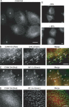
Figure 2. Immunolocalization of CVAK104.
A) HeLa cells were either treated with siRNA against CVAK104 or untreated (control), then mixed and labelled with anti-CVAK104 antibody. Asterisks indicate CVAK104-depleted cells. Scale bar: 10 μm. B–D) Double labelling for CVAK104 and coat proteins. B) HeLa cells were double labelled for CVAK104 and CHC. The two patterns show significant colocalization. C) HeLa cells were double labelled for CVAK104 and AP-1 γ. Again the two patterns show significant colocalization. D) HeLa cells were double labelled for CVAK104 and AP-2 α. The two patterns show no significant colocalization. Scale bars: 5 μm. E) HeLa cells were treated with BFA (20 μg/mL) for 5 minutes at 37 °C or treated with medium only (control), and labelled with anti-CVAK104 antibody. Upon BFA treatment, CVAK104 loses its membrane association and becomes largely cytosolic. Scale bars: 10 μm. CHC, clathrin heavy chain. Arrowheads indicate areas of colocalisation.
Figure 3
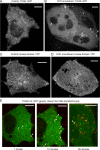
Figure 3. Live-cell imaging of tagged CVAK104 constructs.
A and B) CVAK104 was tagged with GFP at the C-terminus and transiently expressed in HeLa cells. A) In control cells, CVAK104-GFP is present in small and highly mobile compartments. B) In clathrin-depleted cells, CVAK104-GFP is more cytosolic but also labels largely immobile vesicular compartments as well as mobile tubular structures (white arrows). C and D) The N-terminal kinase-like domain of CVAK104 was tagged with YFP and transiently expressed in HeLa cells. C) In control cells, the construct localizes to mobile tubular and vesicular structures. D) In clathrin-depleted cells, the construct localizes mainly to tubular mobile structures. E) CVAK104-GFP was transiently expressed in HeLa cells. Cells were allowed to endocytose pre-bound Alexa Fluor-labelled transferrin, and imaged either ∼1, ∼10 or ∼60 minutes after beginning of uptake by spinning-disk confocal microscopy. Frequent colocalization (white arrows) can be observed at early, but not at late stages of the experiment. Time-points 1 and 10 minutes show images of the same cell, taken from a continuous movie (Video S5). Representative still images are shown here; the corresponding movies are available as Videos S1–S6 (online supplemental material). Scale bars: 10 μm.
Figure 4
Figure 4. CVAK104 kd results in specific depletion of syntaxin 8 and vti1b from CCVs.
Cell homogenates and CCV fractions were prepared from control and siRNA-treated cells and analysed by Western blot. CVAK kd1 and CVAK kd2 denote samples from two independent kd experiments. Approximately 10 μg protein of whole cell lysates or 2.5 μg of CCV fractions were loaded in each lane. Although the four SNAREs, syntaxin 7, syntaxin 8, vti1a, and vti1b, are all sorted by clathrin, only syntaxin 8 and vti1b are depleted from CCVs in a CVAK104 kd. CHC, clathrin heavy chain; kd, knockdown.
Figure 5
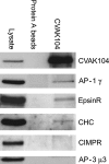
Figure 5. CVAK104 interacts with clathrin, AP-1 and epsinR.
HeLa cells were extracted with 1% digitonin and immunoprecipitated with an antibody against CVAK104 in conjunction with protein A linked to Sepharose beads, or with protein A beads only (control). Immunoprecipitates were analysed by Western blot. CHC, clathrin heavy chain.
Figure 6
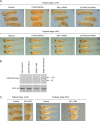
Figure 6. CVAK104 knockdown and rescue in Xenopus.
A) Fertilized X. tropicalis eggs were injected with a control MO (panels 1 and 5) or a MO against CVAK104 (panels 2 and 6) at the one-cell stage and imaged either 24 h or 48 h post-fertilization. To rescue the knockdown phenotype, fertilized eggs were injected with anti-CVAK104 MO + mRNA encoding human CVAK104-myc (panels 3 and 7), then imaged either 24 h or 48 h post-fertilization. Expression of human CVAK104-myc resulted in high levels of rescue both at tailbud and tadpole stages. Expression of human CVAK104-myc alone had no apparent detrimental effects (panels 4 and 8). B) Western blots of extracts from 24 h embryos were probed with anti-myc antibody. AP-2 μ2 was included as a loading control. Human CVAK104-myc was detectable in embryos that had been injected with mRNA. C) Fertilized eggs were injected with either a control MO, a MO against AP-2 μ2, or a MO against AP-1 μ1A, then imaged either 24 h or 48 h post-fertilization. Both knockdowns cause severe phenotypes, but the AP-1 knockdown phenotype is more similar to the CVAK104 knockdown phenotype.

References

    1. Robinson MS. Adaptable adaptors for coated vesicles. Trends Cell Biol. 2004;14:167–174. - PubMed
    1. Edeling MA. Life of a clathrin coat: insights from clathrin and AP structures. Nat Rev Mol Cell Biol. 2006;7:32–44. - PubMed
    1. Traub LM. Common principles in clathrin-mediated sorting at the Golgi and the plasma membrane. Biochim Biophys Acta. 2005;1744:415–437. - PubMed
    1. Bonifacino JS. Retrograde transport from endosomes to the trans-Golgi network. Nat Rev Mol Cell Biol. 2006;7:568–579. - PubMed
    1. Foote C. The clathrin adaptor complex 1 directly binds to a sorting signal in Ste13p to reduce the rate of its trafficking to the late endosome of yeast. J Cell Biol. 2006;173:615–626. - PMC - PubMed

Publication types