

Optical biosensor differentiates signaling of endogenous PAR1 and PAR2 in A431 cells
- PMID: 17587449
- PMCID: PMC1925066
- DOI: 10.1186/1471-2121-8-24
Optical biosensor differentiates signaling of endogenous PAR1 and PAR2 in A431 cells
Abstract
Background: Protease activated receptors (PARs) consist of a family of four G protein-coupled receptors. Many types of cells express several PARs, whose physiological significance is mostly unknown.
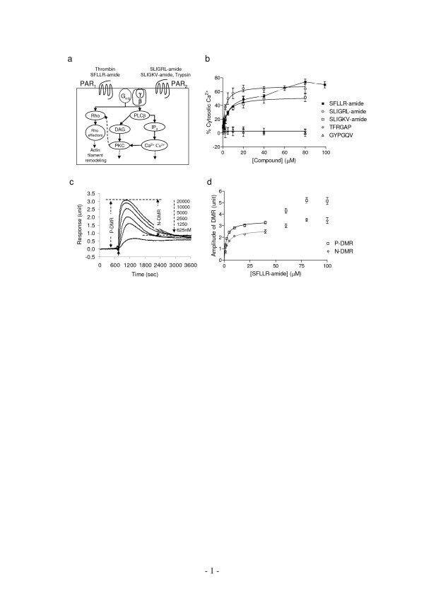
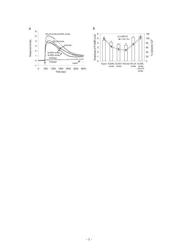
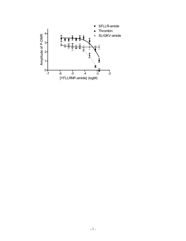
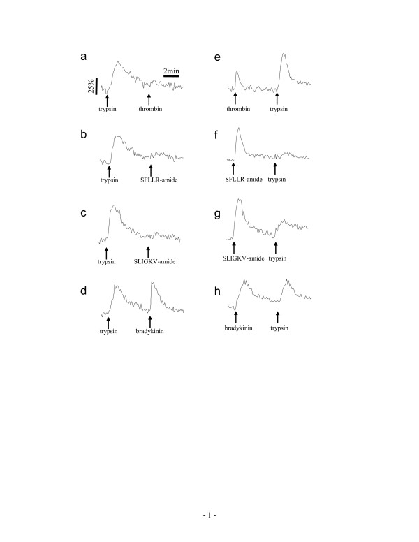
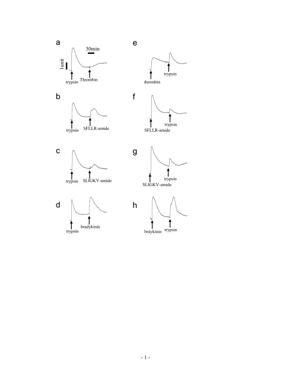
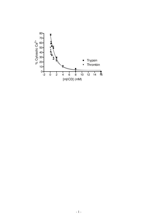
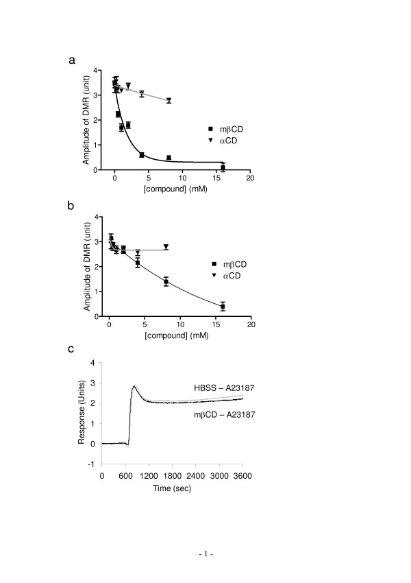
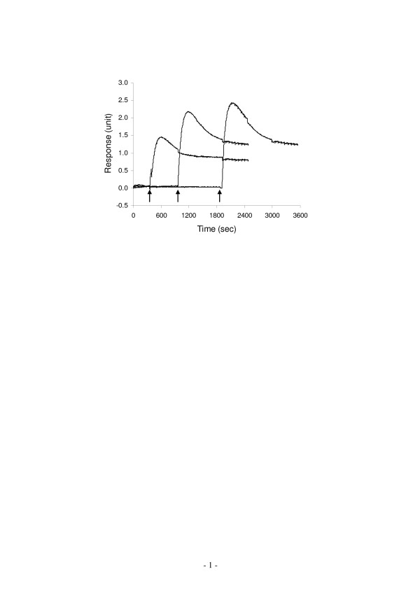
Results: Here, we show that non-invasive resonant waveguide grating (RWG) biosensor differentiates signaling of endogenous protease activated receptor subtype 1 (PAR1) and 2 (PAR2) in human epidermoid carcinoma A431 cells. The biosensor directly measures dynamic mass redistribution (DMR) resulted from ligand-induced receptor activation in adherent cells. In A431, both PAR1 and PAR2 agonists, but neither PAR3 nor PAR4 agonists, trigger dose-dependent Ca2+ mobilization as well as Gq-type DMR signals. Both Ca2+ flux and DMR signals display comparable desensitization patterns upon repeated stimulation with different combinations of agonists. However, PAR1 and PAR2 exhibit distinct kinetics of receptor re-sensitization. Furthermore, both trypsin- and thrombin-induced Ca2+ flux signals show almost identical dependence on cell surface cholesterol level, but their corresponding DMR signals present different sensitivities.
Conclusion: Optical biosensor provides an alternative readout for examining receptor activation under physiologically relevant conditions, and differentiates the signaling of endogenous PAR1 and PAR2 in A431.
Figures

References
-
- Negrescu EV, de Quintana KL, Siess W. Platelet shape change induced by thrombin receptor activation. Rapid stimulation of tyrosine phosphorylation of novel protein substrates through an integrin- and Ca2+-independent mechanism. J Biol Chem. 1995;270:1057–1061. doi: 10.1074/jbc.270.3.1057. - DOI - PubMed
MeSH terms
Substances
LinkOut - more resources
Full Text Sources
Other Literature Sources
Miscellaneous
