. 2007 Jun 24:7:10.
doi: 10.1186/1471-2482-7-10.

Hepatobiliary and pancreatic tuberculosis: a two decade experience

Affiliations

Hepatobiliary and pancreatic tuberculosis: a two decade experience

Sundeep S Saluja et al. BMC Surg. .

Abstract

Background: Isolated hepatobiliary or pancreatic tuberculosis (TB) is rare and preoperative diagnosis is difficult. We reviewed our experience over a period two decades with this rare site of abdominal tuberculosis.

Methods: The records of 18 patients with proven histological diagnosis of hepatobiliary and pancreatic tuberculosis were reviewed retrospectively. The demographic features, sign and symptoms, imaging, cytology/histopathology, procedures performed, outcome and follow up data were obtained from the departmental records. The diagnosis of tuberculosis was based on granuloma with caseation necrosis on histopathology or presence of acid fast bacilli.

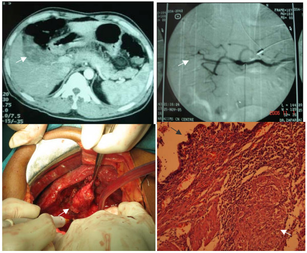
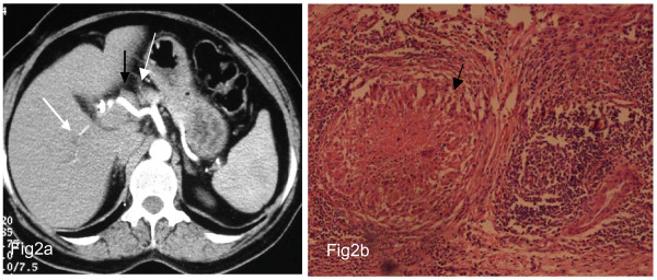
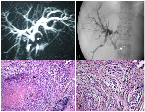
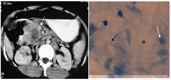
Results: Of 18 patients (11 men), 11 had hepatobiliary TB while 7 had pancreatic TB. Two-thirds of the patients were < 40 years (mean: 42 yrs; range 19-70 yrs). The duration of the symptoms varied between 2 weeks to 104 weeks (mean: 20 weeks). The most common symptom was pain in the abdomen (n = 13), followed by jaundice (n = 10), fever, anorexia and weight loss (n = 9). Five patients (28%) had associated extra-abdominal TB which helped in preoperative diagnosis in 3 patients. Imaging demonstrated extrahepatic bile duct obstruction in the patients with jaundice and in addition picked up liver, gallbladder and pancreatic masses with or without lymphadenopathy (peripancreatic/periportal). Preoperative diagnosis was made in 4 patients and the other 14 were diagnosed after surgery. Two patients developed significant postoperative complications (pancreaticojejunostomy leak 1 intraabdominal abscess 1) and 3 developed ATT induced hepatotoxicity. No patient died. The median follow up period was 12 months (9-96 months).

Conclusion: Tuberculosis should be considered as a differential diagnosis, particularly in young patients, with atypical signs and symptoms coming from areas where tuberculosis is endemic and preoperative tissue and/or cytological diagnosis should be attempted before labeling them as hepatobiliary and pancreatic malignancy.


Figures

Figure 1
Figure 1
Case 1 A 41-year-old man presented with upper gastrointestinal bleed. Endoscopy revealed blood coming out from ampulla Fig 1a Axial section of Dual phase CT scan shows hyperdensity (white arrow) within the gallbladder 1b Flush aortogram shows origin of the right hepatic artery directly from the coeliac trunk and spasm in one of the branches (white arrow) 1 c Intraoperative picture shows multiple tubercles over the gallbladder (white arrow) and liver surface 1 d Histopathology sections (100×) showing gallbladder mucosa (black arrow) with underlying epithelioid cell granuloma (white arrow). Final diagnosis: Gall bladder TB
Figure 2
Figure 2
Case 2 A 55-year-old woman presented with obstructive jaundice and cholangitis for 2 months. Past history of cholecystectomy done 1 year ago. Fig 2a CECT showed mildly dilated intrahepatic radicals (white arrow) with enlarged lymph nodes (black arrow) along the hepatic artery. On exploration mass in CBD with multiple pericholedochal lymph nodes seen. Frozen section of the lymph node showed nectrotizing granuloma. 2b) Histopathology sections (40×) show epithelioid cell granuloma with central area of caseous necrosis (white arrow). Final diagnosis: Bile duct TB secondary to periportal lymph nodes
Figure 3
Figure 3
Case 3 A 56-year-old woman presented with obstructive jaundice (2 months) associated with cholangitis and previous cholecystectomy (8 years ago). Fig 3 a) MRCP done showed dilated intrahepatic radicals with stricture below confluence (white arrow). 3 b) PTBD done to relieve cholangitis shows stricture below confluence (white arrow). At exploration 2 × 1 cm mass in CBD encasing portal vein with multiple pericholedochal lymph nodes. She underwent palliative hepaticojejunostomy and biopsy of the bile duct and lymph node 3 c) Histopathology sections examined under low power (40×) show epitheloid cell granuloma with central area of caseous necrosis (black arrow). At the periphery multiple ducts are seen with periductal chronic inflammation and fibrosis. 3 d) Sections examined under high power (200×) show ducts with dense chronic inflammatory infiltrates comprising of lymphocytes along with periductal fibrosis (black arrow). Final diagnosis: Biliary tuberculosis
Figure 4
Figure 4
Case 4 A 23-year-old woman presented with pain abdomen, weight loss and enlarged supraclavicular lymph nodes for 2 years. Fig 4a Axial section of CECT abdomen shows 4 × 2 cm hypodense mass in the region of the head of the pancreas (white arrow) with multiple peripancreatic lymph nodes. 4b) FNAC from the mass showed acid-fast bacilli (black arrow) surrounded by epithelioid cells (white arrow) Final diagnosis: Pancreatic tuberculosis

Similar articles

Cited by

References

    1. Leder RA, Low VHS. Tuberculosis of the abdomen. Radiol Clin N Am. 1995;33:691–705. - PubMed
    1. Chen C, Yang C, Yeh Y, Yang J, Chou D. Pancreatic tuberculosis with obstructive jaundice – A case report. Am J Gastroenterol. 1999;94:2534–253. - PubMed
    1. Hulnick DH, Megibow AJ, Naidich DP, Hilton S, Cho KC, Balthazar EJ. Abdominal tuberculosis: CT evaluation. Radiology. 1985;157:199–204. - PubMed
    1. Aston NO, Chir M. Abdominal tuberculosis. World J Surg. 1997;24:492–499. doi: 10.1007/PL00012275. - DOI - PubMed
    1. Sinan T, Sheikh M, Ramadan S, Sahwney S, Behbehani A. CT features in abdominal tuberculosis: 20 years experience. BMC Med Imaging. 2002;2:3–10. doi: 10.1186/1471-2342-2-3. - DOI - PMC - PubMed