Review
. 2008 Jan 1;469(1):100-17.
doi: 10.1016/j.abb.2007.05.015. Epub 2007 Jun 8.

Folding versus aggregation: polypeptide conformations on competing pathways

Affiliations
Review

Folding versus aggregation: polypeptide conformations on competing pathways

Thomas R Jahn et al. Arch Biochem Biophys. .

Abstract

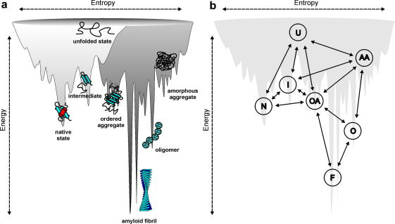
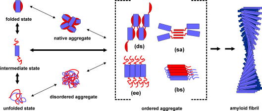
Protein aggregation has now become recognised as an important and generic aspect of protein energy landscapes. Since the discovery that numerous human diseases are caused by protein aggregation, the biophysical characterisation of misfolded states and their aggregation mechanisms has received increased attention. Utilising experimental techniques and computational approaches established for the analysis of protein folding reactions has ensured rapid advances in the study of pathways leading to amyloid fibrils and amyloid-related aggregates. Here we describe recent experimental and theoretical advances in the elucidation of the conformational properties of dynamic, heterogeneous and/or insoluble protein ensembles populated on complex, multidimensional protein energy landscapes. We discuss current understanding of aggregation mechanisms in this context and describe how the synergy between biochemical, biophysical and cell-biological experiments are beginning to provide detailed insights into the partitioning of non-native species between protein folding and aggregation pathways.


Figures

Fig. 1
Fig. 1
Illustration of a combined energy landscape for protein folding and aggregation. (a) The surface illustrates the roughness of the protein energy landscape, showing the multitude of conformational states available to a polypeptide chain. While rather simple folding funnels (light grey) can describe the conformational search of a single polypeptide chain to a functional monomer, intermolecular protein association dramatically increases ruggedness (dark grey). (b) Proposed pathways linking the conformational states shown in (a) populated on the combined folding and aggregation energy landscape.
Fig. 2
Fig. 2
Application of restrained MD simulations to describe the conformational properties of dynamic ensembles. Structural ensembles of CI2 corresponding to (a) the crystal structure of the native state, (b) the native state ensemble determined from hydrogen exchange data and (c) the TSE determined using experimental phi-values. (d) Comparison of experimental protection factors (black circles) with those back-calculated (solid lines) from the native state ensemble shown in (b). (e) Agreement between the phi-values calculated from the TSE ensemble shown in c (black line) and experimental phi-values (red circles). Figure adapted from with permission.
Fig. 3
Fig. 3
Proposed mechanisms of protein aggregation. Amyloid fibril formation for many proteins proceeds from intermediate partially folded states that are formed via partial unfolding of the native structure or via the partial structuring of unfolded polypeptides. Ordered aggregates associate via mechanisms such as domain swapping (ds), strand association (sa), edge-edge-association (ee) or β-strand stacking (bs). Self-association of these early oligomeric species, possibly involving further conformational changes, then leads to the formation of amyloid fibrils. The generic principles that govern this self-association process and the structure of the final amyloid fibril may depend critically on the polypeptide sequence and the solution conditions. In some proteins association of native-like monomers or non-specific self-association into disordered aggregates has been observed as the initial step in amyloid assembly, in the latter route the polypeptide adopts an ordered β-sheet structure within the initially disordered aggregate before amyloid fibril formation proceeds.
Fig. 4
Fig. 4
Pathway complexity of β2m amyloid fibril formation. (a) Schematic state diagram representing the different thermodynamic ground states observed upon incubation of β2m under different conditions. While the native protein (N) remains monomeric even at high protein concentration, acidification results in the protein unfolding to form partially folded (PF) or more highly unfolded forms (U). Above a critical concentration these species self-associate, forming amyloid fibrils with distinct morphological properties. Close to the protein’s pI, amorphous aggregates (AA) are formed, while worm-like fibrils (WL) and classic long-straight fibrils (LS) are formed at lower pH values. (b) Proposed model for competing pathways that lead to the formation of worm-like fibrils or long-straight amyloid fibrils. (c,d) AFM images of worm-like (c) and long-straight fibrils (d). All images are 1 μm2. (e,f) Worm-like fibrils form with nucleation-independent kinetics (e), whilst the formation of long-straight fibrils is nucleation-dependent and shows a clear lag-phase (f). Figure adapted from with permission.
Fig. 5
Fig. 5
General structural motifs of amyloid-like fibrils. (a–c) Structural model for protofilaments formed in vitro by a WW domain. (a) Perpendicular view to the fibril axis, indicating the continuous β-sheet hydrogen bond structure (dotted lines). (b) Cartoon representation of the non-native β-strand-loop-β-strand motif adopted in the amyloid structure. Structural restraints from ssNMR measurements are indicated by arrows and side chains eliminating (red) or reducing (orange) amyloid fibril formation when mutated to alanine are highlighted. (c) Atomistic representation of the tight packing between strands (viewed along the fibril axis as in (b)). (d–g) Surface representation of 3D maps obtained using cryoEM for insulin fibril structures. The fibrils contain either two (d), four (e) or six (f and g) protofilaments. (h) Proposed β-strand model for insulin fibrils containing four protofilaments. Figure adapted from references and with permission.
Fig. 6
Fig. 6
Representative structures of proteins involved in disease-related amyloid fibril formation. The polypeptides are coloured according to the aggregation tendency of their amino acid sequences predicted using the algorithm TANGO . Sequences shown in blue are predicted to have no β-aggregation propensity, while polypeptide stretches coloured in yellow, orange and red indicate an increasing propensity to aggregate. Notably, the peptide structures were obtained in the presence of fluoroalcohols (calcitonin and Aβ1–42) or SDS micelles (amylin), and these sequences might be substantially less ordered in the absence of these additives. Note also that for insulin, amylin and calcitonin, the pro-peptides as well as the mature sequences have been implicated as potentially amyloidogenic [225–227].
Fig. 7
Fig. 7
Schematic illustration of the importance of edge-strands in the aggregation of β-sheet proteins. Local unfolding of the protective edge-strands (orange) has been implicated in amyloid fibril formation for (a) β2m using NMR spectroscopy , (b) TTR using hydrogen exchange experiments and (c) SOD using protein engineering .

References

    1. Levinthal C. J. Chim. Phys. 1968;65:44–45.
    1. Fersht A.R., Daggett V. Cell. 2002;108:573–582. - PubMed
    1. Dinner A.R., Sali A., Smith L.J., Dobson C.M., Karplus M. Trends Biochem. Sci. 2000;25:331–339. - PubMed
    1. Wolynes P.G., Onuchic J.N., Thirumalai D. Science. 1995;267:1619–1620. - PubMed
    1. Dill K.A., Chan H.S. Nature Struct. Biol. 1997;4:10–19. - PubMed

Publication types

LinkOut - more resources