Review
. 2007 Aug-Sep;28(2-3):97-114.
doi: 10.1016/j.yfrne.2007.04.002. Epub 2007 May 21.

Negative feedback regulation of hypophysiotropic thyrotropin-releasing hormone (TRH) synthesizing neurons: role of neuronal afferents and type 2 deiodinase

Affiliations
Review

Negative feedback regulation of hypophysiotropic thyrotropin-releasing hormone (TRH) synthesizing neurons: role of neuronal afferents and type 2 deiodinase

Csaba Fekete et al. Front Neuroendocrinol. 2007 Aug-Sep.

Abstract

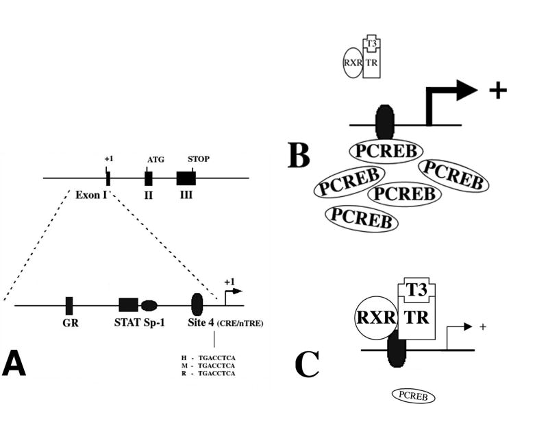
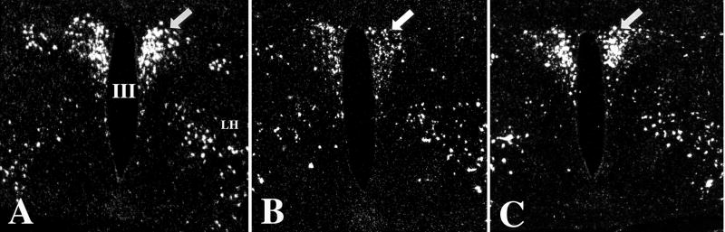
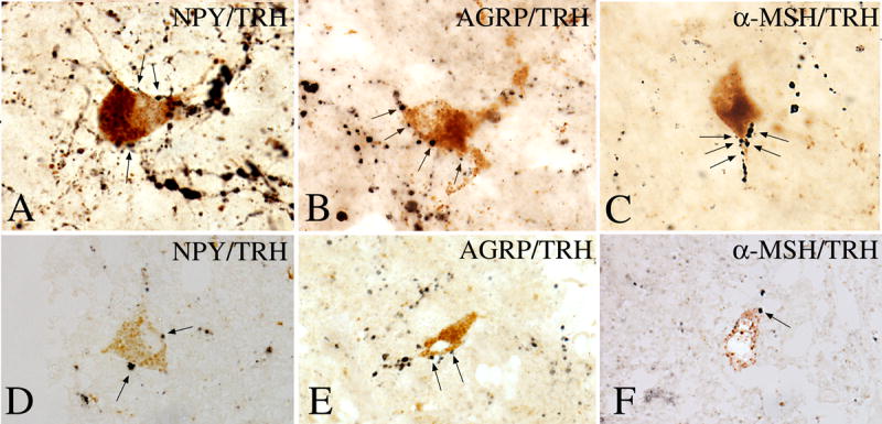
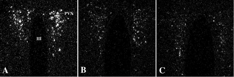
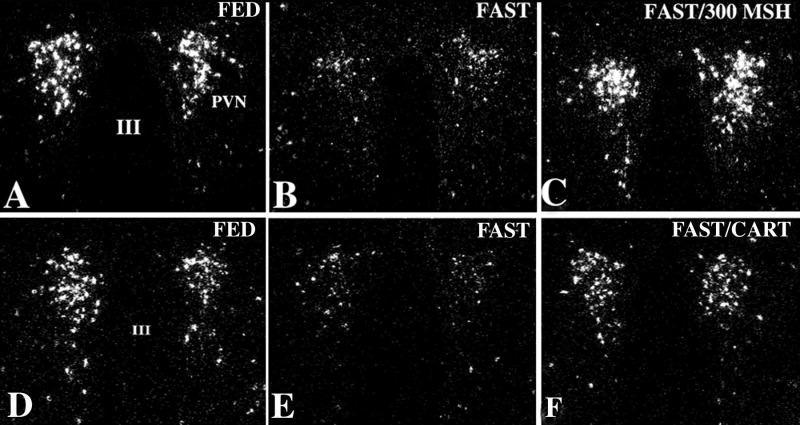
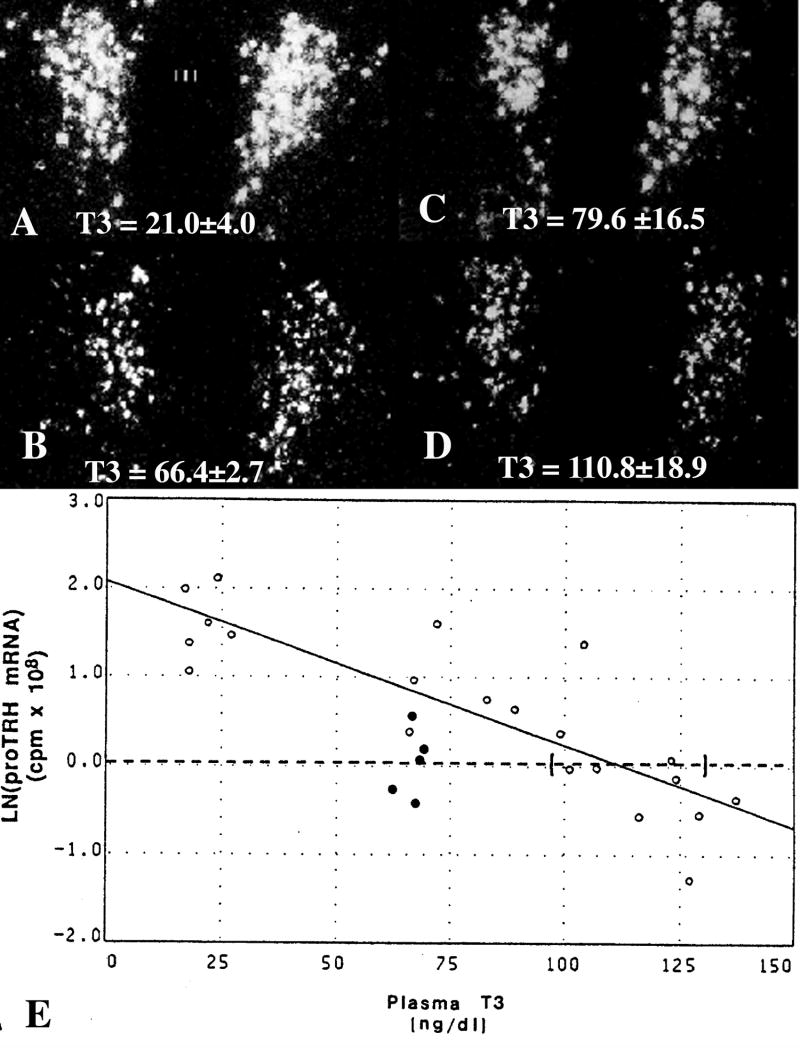
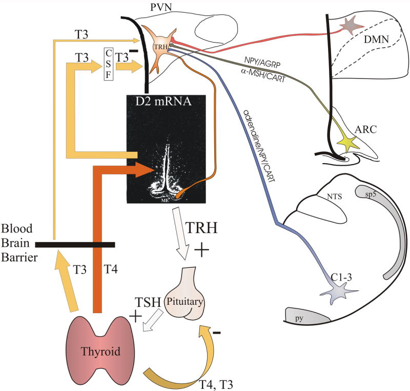
Hypophysiotropic thyrotropin-releasing hormone (TRH): synthesizing neurons reside in the hypothalamic paraventricular nucleus (PVN) and are the central regulators of the hypothalamic-pituitary-thyroid (HPT) axis. TRH synthesis and release from these neurons are primarily under negative feedback regulation by thyroid hormone. Under certain conditions such as cold exposure and fasting, however, inputs from neurons in the brainstem and hypothalamic arcuate and dorsomedial nuclei alter the set point for negative feedback through regulation of CREB phosphorylation. Thus, during cold exposure, adrenergic neurons stimulate the HPT axis, while fasting-induced central hypothyroidism is mediated through an arcuato-paraventricular pathway. Feedback regulation of TRH neurons may also be modified by local tissue levels of thyroid hormone regulated by the activation of type 2 iodothyronine deiodinase (D2), the primary enzyme in the brain that catalyzes T4 to T3 conversion. During infection, endotoxin or endotoxin induced cytokines increase D2 activity in the mediobasal hypothalamus, which by inducing local hyperthyroidism, may play an important role in infection-induced inhibition of hypophysiotropic TRH neurons.


Figures

Figure 1
Figure 1
Distribution of TRH-synthesizing neurons in the PVN. Low power micrographs (A–C) illustrate the TRH neurons at three rostrocaudal levels of the PVN. Schematic drawings (D–F) illustrate the subdivisions of the PVN where hypophysiotropic TRH neurons are localized (gray). AP, anterior parvocellular subdivision; DP, dorsal parvocellular subdivision; LP, lateral parvocellular subdivision; MN, Magnocellular part of PVN; MP, medial parvocellular subdivision, PV, periventricular parvocellular subdivision, III, third ventricle
Figure 2
Figure 2
Distribution of TRH-immunoreactive (IR) terminals in the median eminence. While TRH-IR axons densely innervate the external zone of the median eminence, only scattered fibers can be seen in the internal zone. III, third ventricle; ME, median eminence
Figure 3
Figure 3
In situ hybridization autoradiograms showing the effect of hypo- and hyperthyroidism on TRH mRNA level in the medial parvocellular subdivision of the PVN. A substantial increase in silver grain accumulation is observed in the hypothyroid animal (B) compared to the fed control (A). In contrast, hyperthyroidism results in a marked reduction of TRH mRNA level in the PVN (C). (From Dyess et al [23], copyright 1988, with permission from The Endocrine Society.)
Figure 4
Figure 4
Presence of TRβ2 isoform (A) in the TRH neurons (B) in the PVN. Arrows indicate the neurons containing both TRβ2 and TRH. (From Lechan et al [22], copyright 1994, with permission from The Endocrine Society.)
Figure 5
Figure 5
Genomic and promoter structure of TRH. The murine, rat and human TRH genes are composed of three exons and two introns. The coding sequence for the precursor protein is present on exons 2 and 3. As depicted, the TRH promoter region precedes the transcription start site in exon 1. The proximal 250 bp sequences of the human, mouse and rat promoters are similar and share the indicated transcription factor binding sites. The location of the CREB binding site (Site 4) and sequences in human (H), mouse (M) and rat (R) are shown. (B and C) Hypothesized schematic representation of the interaction between PCREB and the thyroid hormone receptor at Site 4. (B) illustrates that in the presence of abundant PCREB, there may be less availability for binding of the thyroid hormone receptor/T3 complex, hence, an increase in TRH gene transcription. When PCREB concentrations fall as shown in (C), increased binding of the thyroid hormone receptor/T3 complex reduces TRH gene transcription. (From Lechan and Fekete [62], copyright 2006, with permission from Elsevier.)
Figure 6
Figure 6
Darkfield illumination photomicrographs of proTRH mRNA in the hypothalamic PVN in fed (A), fasted (B), and fasted animals receiving leptin (C). Note the marked reduction in silver grains over neurons in the PVN in the fasted animals but restoration to normal in the fasted animals receiving leptin. III, third ventricle (From Legradi et al, copyright 1997 [36], with permission from The Endocrine Society.)
Figure 7
Figure 7
The association between axon terminals containing NPY, AGRP, and α-MSH-immunoreactivity (arrows) with TRH-positive perikarya in the PVN of human hypothalamus is shown in A–F. A multipolar TRH-IR neuron is heavily contacted by NPY-IR axon terminals (A). AGRP-IR axon varicosities establish numerous axo-somatic and axo-dendritic connections with a TRH-positive cell (B). α-MSH-IR axon varicosities densely cover the dendrite of a TRH-containing neuron (C). Similar associations between NPY-, AGRP-, and α-MSH-IR axon terminals and TRH-containing cells are shown in 1-μm thick sections in D–F. (From Mihály et al [160], copyright 2000, with permission from The Endocrine Society.)
Figure 8
Figure 8
Darkfield illumination photomicrographs of proTRH mRNA in the medial parvocellular subdivision of the PVN in control (A), AGRP- (B), and NPY-treated (C) animals. Note marked reduction in silver grains over neurons in the PVN in both the AGRP- and NPY-infused groups. III, Third ventricle (From Fekete et al [76], copyright 2002, with permission from The Endocrine Society.)
Figure 9
Figure 9
Dark-field illumination micrographs of pro-TRH mRNA in the medial and periventricular parvocellular subdivisions of the hypothalamic PVN in fed (A, D) and fasted (B, E) animals and fasted animals receiving an intracerebroventricular infusion of α-MSH (C) or CART (F) every 6 hr for 64 hr. Note the reduction in the accumulation of silver grains over the PVN in fasted animals compared with the fed controls. Both α-MSH and CART administration prevent the fasting induced fall of TRH mRNA levels. III, Third ventricle (From Fekete et al, copyright 2000, with permission from The Endocrine Society.)
Figure 10
Figure 10
In situ hybridization autoradiographs of proTRH mRNA in the paraventricular nucleus (PVN) in (A) hypothyroid, (B) euthyroid and (C) hypothyroid animals receiving a constant infusion of (C) 0.5 μg or (D) 0.75 μg of T3/100 gm/bw/d. Mean plasma triiodothyronine (T3) levels (±SEM) are shown for each group at the bottom of the photomicrographs. Note that only the higher dose of T3 that raised plasma T3 levels into the supranormal range was capable of suppressing TRH mRNA to euthyroid levels. (E) Regression analysis of above experiment. Interrupted line represents the mean ln(TRH mRNA) for euthyroid animals and its intercept with the regression line estimates the plasma T3 concentration required to suppress proTRH mRNA to euthyroid levels. Ninety-five percent confidence intervals for each intercept are bracketed. Open dots denote values for hypothyroid animals and hypothyroid animals infused with graded doses of T3. Closed dots denote values for euthyroid controls. (Modified from Kakucska et al [109], copyright 1992, with permission from The Endocrine Society.)
Figure 11
Figure 11
Distribution of type 2 deiodinase mRNA in tanycytes lining the wall of the third ventricle. Low power micrographs of a rostral (A) and a caudal (B) section show that silver grains denoting type 2 deiodinase mRNA are accumulated over the cells lining the wall of the third ventricle, the tuberoinfundibular sulci (arrow heads) and around blood vessels in the arcuate nucleus (arrows). Note the absence of the hybridization signal in the upper third of the ventricle (open arrow). Higher power micrographs shows the association of the silver grains with the tuberoinfundibular sulcus (arrow heads) and a blood vessel (arrows) in the arcuate nucleus (C), and the presence of type 2 deiodinase mRNA in the external zone of the median eminence (D). (Modified from Fekete et al [118], copyright 2000, with permission from Elsevier.)
Figure 12
Figure 12
Low-power dark-field micrographs from two different rostrocaudal levels of the median eminence (ME) showing the effect of LPS treatment on D2 gene expression in the MBH. A–B, Controls; C–D, LPS-treated animals. Silver grains denoting D2 mRNA are accumulated over cells lining the wall of the third ventricle (III), the tuberoinfundibular sulci (arrow heads), and accumulate in the external zone of the ME. After LPS administration, the density of silver grains denoting D2 mRNA is markedly increased, particularly in the external zone of the ME (C–D). (Modified from Fekete et al [146], copyright 2004, with permission from The Endocrine Society.)
Figure 13
Figure 13
Schematic illustration of the feedback system regulating the hypothalamic-pituitary-thyroid axis. Thyroid hormones exert negative feedback effect at the level of the pituitary and hypophysiotropic TRH neurons. The central feedback effect of thyroid hormones primarily depends on the circulating T4 levels. In the hypothalamus, T4 is converted to T3 by D2 in tanycytes. By volume transmission, T3 secreted from tanycytes reaches the hypophysiotropic TRH neurons, where T3 inhibits the proTRH gene expression via TRβ2 receptors. The setpoint of the feedback regulation can be altered by two mechanisms: 1) Regulation of D2 activity in tanycytes may alter the hypothalamic T3 availability independently from the peripheral T4 concentration. 2) Neuronal afferents can alter the PCREB concentration in the hypophysiotropic TRH neurons that can change the setpoint of feedback regulation through competition of PCREB and thyroid hormone receptors for the multifunctional binding site (Site 4) of the TRH promoter. ARC, hypothalamic arcuate nucleus; C1-3, C1-3 adrenergic area of the brainstem; CSF, cerebrospinal fluid; DMN, hypothalamic dorsomedial nucleus; ME, median eminence; NTS, nucleus tractus solitarius; PVN, hypothalamic paraventricular nucleus; py, pyramidal tract; sp5, spinal trigeminal tract

References

    1. Lechan RM. Update on thyrotropin-releasing hormone. Thyroid Today. 1993;16:1–12.
    1. Nillni EA, Sevarino KA. The biology of prothyrotropin-releasing hormone-derived peptides. Endocr Rev. 1999;20:599–648. - PubMed
    1. Bradbury AF, Finnie MD, Smyth DG. Mechanism of C-terminal amide formation by pituitary enzymes. Nature. 1982;298:686–688. - PubMed
    1. Fischer WH, Spiess J. Identification of a mammalian glutaminyl cyclase converting glutaminyl into pyroglutamyl peptides. Proc Natl Acad Sci U S A. 1987;84:3628–3632. - PMC - PubMed
    1. Lechan RM, Segerson TP. Pro-TRH gene expression and precursor peptides in rat brain. Observations by hybridization analysis and immunocytochemistry. Ann N Y Acad Sci. 1989;553:29–59. - PubMed

Publication types

MeSH terms

Substances