Review
. 2007 Aug 29;362(1484):1369-91.
doi: 10.1098/rstb.2007.2122.

Heart valve function: a biomechanical perspective

Affiliations
Review

Heart valve function: a biomechanical perspective

Michael S Sacks et al. Philos Trans R Soc Lond B Biol Sci. .

Erratum in

  • Philos Trans R Soc Lond B Biol Sci. 2008 Jul 27;363(1502):2471

Abstract

Heart valves (HVs) are cardiac structures whose physiological function is to ensure directed blood flow through the heart over the cardiac cycle. While primarily passive structures that are driven by forces exerted by the surrounding blood and heart, this description does not adequately describe their elegant and complex biomechanical function. Moreover, they must replicate their cyclic function over an entire lifetime, with an estimated total functional demand of least 3x10(9) cycles. As in many physiological systems, one can approach HV biomechanics from a multi-length-scale approach, since mechanical stimuli occur and have biological impact at the organ, tissue and cellular scales. The present review focuses on the functional biomechanics of HVs. Specifically, we refer to the unique aspects of valvular function, and how the mechanical and mechanobiological behaviours of the constituent biological materials (e.g. extracellular matrix proteins and cells) achieve this remarkable feat. While we focus on the work from the authors' respective laboratories, the works of most investigators known to the authors have been included whenever appropriate. We conclude with a summary and underscore important future trends.


Figures

Figure 1
Figure 1
A schematic of the role of biomechanics in HV function, which occurs at multiple structural levels. At each structural level, biomechanics plays a functionally critical role. This is especially true at the cell and tissue levels, which will control functional equivalence and determine long-term growth/durability.
Figure 2
Figure 2
Numerical simulation of unsteady, pulsatile flow in a tricuspid prosthetic HV. Contours of the out-of-plane vorticity are shown at two instants during the cardiac cycle: (a) fully open phase and (b) closing phase.
Figure 3
Figure 3
Instantaneous friction streamline and shear stress magnitude plots on the aortic (a,c) and ventricular (b,d) sides of the leaflets during the fully open (a,b) and early closing (c,d) phases of the cardiac cycle.
Figure 4
Figure 4
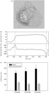
(a) A schematic of the MV anterior leaflet showing the nine transducer sonomicrometry array. (b) Representative time–areal strain traces, along with the corresponding areal strain rate data. Strain rates were quite high, on the order of 1000% s−1, underscoring the highly dynamic nature of the MV. Moreover, once the valve had fully coapted there were no further deformations. (c) Mean principal strains for three pressures levels. Other than the differences between circumferential and mean radial peak strains, there were no significant differences with increasing LV pressure. Adapted from Sacks et al. (2006).
Figure 5
Figure 5
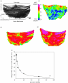
(a) A diagram of the AV cusp showing the locations of the belly, commissure and nodulus, regions of coaptation. SALS results for the AV cusp at (b) 0, (c) 4 and (d) 90 mmHg TVPs. Here, the lines represent preferred collagen direction and colour fringes the local degree of fibre alignment. Past approximately 4 mmHg, no further changes in fibre alignment were observed. This is consistent with histological-based observations were the per cent area of the tissue displaying collagen fibre crimp drops below 10% beyond TVPs of approximately 20 mmHg, as shown in (e). Adapted from Sacks et al. (1998).
Figure 6
Figure 6
(a) Schematic of the biaxial mechanical test configuration for the aortic valve leaflet. (b) Representative biaxial mechanical data taken for the native aortic valve under planar biaxial stretch. Note the large strains and dramatic degree of mechanical anisotropy, with the radial direction exhibiting much larger strains. (c) Simulations using the structural model of the effect of σ on the equibiaxial stress–strain behaviour. The insets provide a graphical representation of the fibre probability density distribution for each σ value: (i) σ=90° approximately isotropic, (ii) σ=35° response qualitatively similar to bovine pericardium, (iii) σ=20° the circumferential strains are negative at low equibiaxial tensions and (iv) σ=10° the material behaviour is highly anisotropic. The dotted lines indicating zero strain are included to highlight the ability of the model to simulate the crossover to negative strain observed in the pressure fixed cusps subjected to equibiaxial tension. Adapted from Billiar & Sacks (2000a,b).
Figure 7
Figure 7
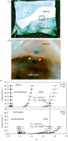
(a) Microdissection of an aortic leaflet specimen, with the test specimen pinned to a cork dissection board such that the ventricularis can be gently lifted to expose the numerous collagen fibre connections coupling the fibrosa and ventricularis. (b) A magnified view of a partially separated leaflet showing the numerous connections found throughout the spongiosa. Each fibrous connection is severed manually enabling us to separate the fibrosa and ventricularis. Note, the markers shown were applied to the outer surface of the fibrosa and ventricularis prior to intact testing and subsequent separation. (c) The equibiaxial responses of the fibrosa and ventricularis computed with respect to both β0 and β1. When referenced to the intact conformation (β0) substantial differences were seen between the radial contributions of each layer. The corresponding first Piola–Kirchhoff stresses were P22v=95.74 kPa while P22f=26.63 kPa at equivalent levels of stretch. These results suggest that the ventricularis layer makes profound contributions to the intact leaflet response in the radial direction. Adapted from Stella & Sacks (in press).
Figure 8
Figure 8
(a) The circumferential and radial stretches of the MV leaflet at the 90 N m−1 equitension state, λCpeak and λRpeak, revealed no significant differences among the prescribed set of loading time protocols in both the circumferential (p=0.987) and radial (p=0.996) directions. Stretches observed previously in mock flow loop (solid horizontal lines) ±s.e.m. (dotted lines) are plotted for the circumferential (black) and radial (grey) specimen axes. λCpeak and λRpeak exceeded those observed in vitro; however, the ratio of λCpeak to λRpeak (0.86±0.02) was very close to the ratio of peak circumferential and radial stretches observed in vitro (0.83). (b) Representative biaxial stress–relaxation data demonstrating continued relaxation throughout the 3 h time frame. (c) Representative stretch versus time curves for a typical biaxial creep experiment. Note the anisotropic leaflet behaviour exhibited by the higher radial stretch required to maintain the 90 N m−1 membrane tension. Only very slight increases (at most 1%) in strain were observed past 1000 s.
Figure 9
Figure 9
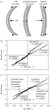
(a) Schematic showing directions of bending for the AV with respective layers (V, ventricularis; S, spongiosa; and F, fibrosa). Note that respective layers are alternating tension and compression resulting from flexural directions. M versus Δκ relations in both the AC and WC directions for (b) specimens tested in 5 mM and 90 mM KCl, and (c) specimens flexed in 5 mM KCl and samples treated in 10 μM thapsigargin overnight and then flexed in 5 mM KCl. While the application of 90 mM KCl induced an increase in stiffness in the AC direction only, both bending directions experienced a loss of stiffness with the addition of thapsigargin to the bathing medium.
Figure 10
Figure 10
Effects of steady shear stress on aortic valve biology. Collagen synthesis and sulphated glycosaminoglycan (sGAG) content in intact (a,b, respectively) and denuded (c,d, respectively) aortic valve leaflets under steady shear stress.
Figure 11
Figure 11
(a) The relation between AVIC NAR and TVP loading, with values reported over the leaflet thickness. Data were taken from native porcine aortic valves fixed from 0 to 90 mmHg pressure. Note clearly that while at 0 mmHg all AVIC have similar NAR, at 90 mmHg the ventricularis layer NAR is approximately 3 while the fibrosa is approximately 4.75. These data indicate that AVICs in the different leaflet layers are subjected to different effective external stresses. (b) AVIC NAR versus the normalized collagen fibre orientation index (NOI) at different TVP levels. Here, we observed that collagen fibre alignment with comparably minimal changes in NAR occur for TVPs up to approximately 4 mmHg. This was followed by the opposite trend for TVPs above 4 mmHg, where minimal changes in NOI were observed accompanied by large changes NAR. These results suggest that AVICs are not appreciably loaded until the collagen fibres fully straighten at TVPs above approximately 4 mmHg.
Figure 12
Figure 12
(a) An example VIC under micropipette aspiration, with the vertical bar indicating aspiration length. (b) Functional correlations of effective cell stiffness E versus TVP, with the correlation coefficient (r) for this relationship of 0.973. (c) Linear correlation between Hsp47 versus SMA, showing a strong correlation between the two proteins (r=0.996) as one progresses from the right to the left side of the heart.

References

    1. Adamczyk M.M, Vesely I. Characteristics of compressive strains in porcine aortic valves cusps. J. Heart Valve Dis. 2002;11:75–83. - PubMed
    1. Akdemir R, Ozhan H, Bulur S, Unlu H, Gunduz H, Arinc H, Yildiz A, Uyan C. Color M-mode regurgitant flow propagation velocity: a new echocardiographic method for grading of mitral regurgitation. Echocardiography. 2005;22:713–722. doi:10.1111/j.1540-8175.2005.00101.x - DOI - PubMed
    1. Arts T, Meerbaum S, Reneman R, Corday E. Stresses in the closed mitral valve: a model study. J. Biomech. 1983;16:539–547. doi:10.1016/0021-9290(83)90068-4 - DOI - PubMed
    1. Bairati A, DeBiasi S. Presence of a smooth muscle system in aortic valve leaflets. Anat. Embryol. 1981;161:329–340. doi:10.1007/BF00301830 - DOI - PubMed
    1. Bellhouse B.J, Bellhouse F.H. Fluid mechanics of the mitral valve. Nature. 1969;224:615–618. doi:10.1038/224615a0 - DOI - PubMed

Publication types

Substances

LinkOut - more resources