Review
. 2007 Aug 29;362(1484):1267-79.
doi: 10.1098/rstb.2007.2114.

Extracellular matrix, mechanotransduction and structural hierarchies in heart tissue engineering

Affiliations
Review

Extracellular matrix, mechanotransduction and structural hierarchies in heart tissue engineering

Kevin K Parker et al. Philos Trans R Soc Lond B Biol Sci. .

Abstract

The spatial and temporal scales of cardiac organogenesis and pathogenesis make engineering of artificial heart tissue a daunting challenge. The temporal scales range from nanosecond conformational changes responsible for ion channel opening to fibrillation which occurs over seconds and can lead to death. Spatial scales range from nanometre pore sizes in membrane channels and gap junctions to the metre length scale of the whole cardiovascular system in a living patient. Synchrony over these scales requires a hierarchy of control mechanisms that are governed by a single common principle: integration of structure and function. To ensure that the function of ion channels and contraction of muscle cells lead to changes in heart chamber volume, an elegant choreography of metabolic, electrical and mechanical events are executed by protein networks composed of extracellular matrix, transmembrane integrin receptors and cytoskeleton which are functionally connected across all size scales. These structural control networks are mechanoresponsive, and they process mechanical and chemical signals in a massively parallel fashion, while also serving as a bidirectional circuit for information flow. This review explores how these hierarchical structural networks regulate the form and function of living cells and tissues, as well as how microfabrication techniques can be used to probe this structural control mechanism that maintains metabolic supply, electrical activation and mechanical pumping of heart muscle. Through this process, we delineate various design principles that may be useful for engineering artificial heart tissue in the future.


Figures

Figure 1
Figure 1
Engineering cell shape and function. Capillary endothelial cells spread to takes on the shape of (a) square (40×40 μm), (b) triangular (long edge 80 μm) and (c) circular (40 μm diameter) ECM islands coated with fibronectin that were created using a microcontact printing technique (Chen et al. 2000), and stained for actin microfilaments using fluorescent-phalloidin. Cells on (a) square and (b) triangular islands preferentially extend motile processes (lamellipodia) from their corners, whereas cells exhibit no bias on the (c) circular islands (Parker et al. 2002; Brock et al. 2003). Note that lamellipodia preferentially extend from acute angles, rather than from the obtuse angle, on the triangle-shaped island.
Figure 2
Figure 2
Control of capillary differentiation using microengineered ECM substrates. (a) Phase contrast and (b) confocal fluorescence microscopic images of capillary endothelial cells forming hollow tubular blood vessels when cultured on fibronectin-coated ECM islands in the form of long thin lines (30 μm wide) that promote only a moderate degree of spreading and support cell–cell contact formation (Dike et al. 1999). A central lumen is visible in both horizontal (left) and vertical (right) cross-sections of the tubes shown in (b).
Figure 3
Figure 3
Coordinated reorganization of the cytoskeleton, focal adhesions and ECM within cells on square islands. Oriented distribution of stress fibres (F-actin), vinculin-containing focal adhesions (vinculin) and underlying fibronectin fibrils (fibronectin) in (a) endothelial cells and (bd) fibroblasts cultured on square fibronectin islands, as detected by fluorescence imaging or atomic force microscopy (AFM). (a) and (b) 50×50 μm islands; (c) and (d) 30×30 μm islands; adapted from Parker et al. (2002).
Figure 4
Figure 4
Geometric control of myofibril architecture in a single cardiomyocyte cultured on a square fibronectin island (50×50 μm). (a) A merged fluorescence microscopic image of the cell after 72 h of culture showing the distribution of sarcomeric α-actinin (red), actin microfilaments (green) and the nucleus (blue). (b) Immunostaining for sarcomeric α-actinin reveals that sarcomerogenesis is initiated in the perinuclear region within the square cell shown on the left. (c) Actin stress fibre bundles that are aligned predominantly along the diagonals of the square in the same cell serve as scaffolds to guide myofibrillogenesis.
Figure 5
Figure 5
The myofibrillar reorganization observed within a single cardiomyocyte cultured on a square fibronectin island (50×50 μm) correlates with the deposition of new fibronectin fibrils in the ECM beneath the cell. (a) Merged image illustrating the position of the cardiomyocyte nucleus (blue), saromeric α-actinin (red) and fibronectin (green). (b) The same cell stained only for sarcomeric α-actinin showing that myofibrillogenesis results in the self-assembly of sarcomeric Z-lines which register and rotate through the internal angles of the corners of the square myocyte, always perpendicular to the cell periphery. (c) Immunostaining for fibronectin reveals fibronectin fibrils beneath the cell in the corners of the square island. This pattern is indicative suggestive of the pattern of tractional forces exerted by the myocyte on the substrate, which are concentrated in these corner regions.
Figure 6
Figure 6
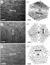
Anisotropic structure–function relations within a microengineered two-dimensional cardiac tissue. Large numbers of cardiomyocytes were cultured on dishes containing a micropatterned array of linear adhesive islands (12.5–25 μm wide) that were oriented in parallel and coated with fibronectin densities of (a) 0, (c) 1.25 or (e)5 μg ml−1. Cells were stained for F-actin using fluorescent-phalloidin to visualize structural anisotropy. Corresponding isochrones of electrical propagation initiated within these tissues by point stimulation at the tissue centre (pulse symbol) are shown at 8 ms intervals in (b), (d) and (f). Double-headed arrows denote direction of micropatterned fibronectin lines and muscle fibre orientation, and the direction of longitudinal propagation of the action potential. (Reprinted with permission from Bursac et al. (2002)).

References

    1. Adler C.P, Ringlage W.P, Bohm N. DNA content and cell number in heart and liver of children. Comparable biochemical, cytophotometric and histological investigations. Pathol. Res. Pract. 1981;172:25–41. [Author's transl.] - PubMed
    1. Alenghat F.J, Ingber D.E. Mechanotransduction: all signals point to cytoskeleton, matrix, and integrins. Sci. STKE. 2002;2002:PE6. - PubMed
    1. Badylak S, Obermiller J, Geddes L, Matheny R. Extracellular matrix for myocardial repair. Heart Surg. Forum. 2003;6:E20–E26. - PubMed
    1. Baicu C.F, Stroud J.D, Livesay V.A, Hapke E, Holder J, Spinale F.G, Zile M.R. Changes in extracellular collagen matrix alter myocardial systolic performance. Am. J. Physiol. Heart Circ. Physiol. 2003;284:H122–H132. - PubMed
    1. Bick R.J, Snuggs M.B, Poindexter B.J, Buja L.M, Van Winkle W.B. Physical, contractile and calcium handling properties of neonatal cardiac myocytes cultured on different matrices. Cell Adhes. Commun. 1998;6:301–310. - PubMed

Publication types

LinkOut - more resources