Comparative Study
. 2007 Sep;25(9):2257-68.
doi: 10.1634/stemcells.2006-0744. Epub 2007 Jun 21.

Markers and methods for cell sorting of human embryonic stem cell-derived neural cell populations

Affiliations
Comparative Study

Markers and methods for cell sorting of human embryonic stem cell-derived neural cell populations

Jan Pruszak et al. Stem Cells. 2007 Sep.

Abstract

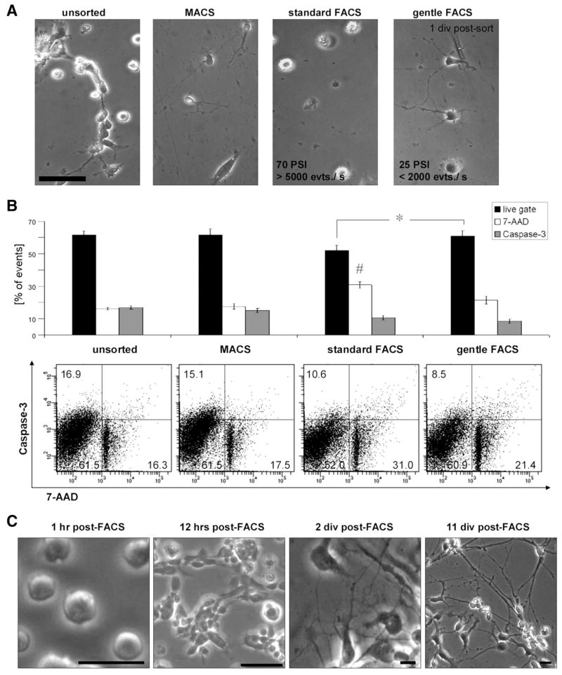
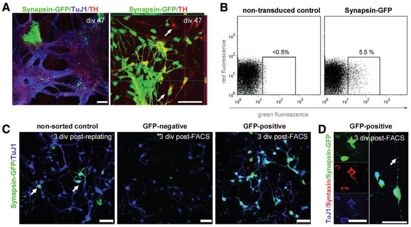
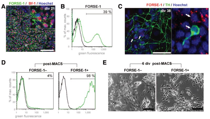
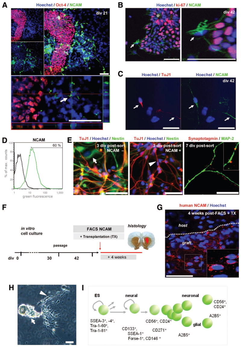
Neural cells differentiated in vitro from human embryonic stem cells (hESC) exhibit broad cellular heterogeneity with respect to developmental stage and lineage specification. Here, we describe standard conditions for the use and discovery of markers for analysis and cell selection of hESC undergoing neuronal differentiation. To generate better-defined cell populations, we established a working protocol for sorting heterogeneous hESC-derived neural cell populations by fluorescence-activated cell sorting (FACS). Using genetically labeled synapsin-green fluorescent protein-positive hESC-derived neurons as a proof of principle, we enriched viable differentiated neurons by FACS. Cell sorting methodology using surface markers was developed, and a comprehensive profiling of surface antigens was obtained for immature embryonic stem cell types (such as stage-specific embryonic antigen [SSEA]-3, -4, TRA-1-81, TRA-1-60), neural stem and precursor cells (such as CD133, SSEA-1 [CD15], A2B5, forebrain surface embryonic antigen-1, CD29, CD146, p75 [CD271]), and differentiated neurons (such as CD24 or neural cell adhesion molecule [NCAM; CD56]). At later stages of neural differentiation, NCAM (CD56) was used to isolate hESC-derived neurons by FACS. Such FACS-sorted hESC-derived neurons survived in vivo after transplantation into rodent brain. These results and concepts provide (a) a feasible approach for experimental cell sorting of differentiated neurons, (b) an initial survey of surface antigens present during neural differentiation of hESC, and (c) a framework for developing cell selection strategies for neural cell-based therapies.


Conflict of interest statement

Disclosure of potential conflicts of interest

The authors indicate no potential conflicts of interest.

Figures

Figure 1
Figure 1
Differentiating hESC are a heterogeneous cell population. (A): Differentiation of hESC in a neuronal induction protocol. In brief, hESC (H1, H7, H9) were neurally induced on Wnt1-MS5 stromal feeder cells with the addition of 300 ng/ml Noggin [2]. Neuroectodermal precursors were then harvested at div 21 and further differentiated using patterning factors such as bFGF, FGF-8, and Shh. For details, see supplemental online methods. (B): During neuronal differentiation in vitro (div 37), continuously proliferative neural precursor cells, in this dopaminergic differentiation paradigm positive for the midbrain-marker Otx-2+, differentiated neuronal cells (TuJ1+), and remaining clusters of immature SSEA-4+stem cells are present. Scale bar: 50 μm. (C): This cellular heterogeneity of hESC differentiation can be illustrated schematically. The developmental potency of immature hESC (t1) may lead to heterogeneity with regard to developmental stage and to cell lineage. The overall population at any given time is therefore composed of different subpopulations, progeny of cells A, B, and C. Remaining immature pluripotent stem and precursor cells may proliferate and spin off progeny at later times (t1–6) and thus increase the anisochronicity of the differentiating cultures. Additionally, non-neural cells, which escape the in vitro patterning factors, develop (supplemental online material 2). Restricting the cultured cells to the population of interest would increase homogeneity and isochronicity of its derivatives for in vitro and in vivo studies. A population purified at an early developmental stage would differentiate free of contamination with unwanted cells more homogenously and synchronize toward the population of interest. Abbreviations: AA, ascorbic acid; BDNF, brain-derived neurotrophic factor; bFGF, basic fibroblast growth factor; cAMP, dibutyryl cyclic adenosine 5′ monophosphate; div, days in vitro; FGF8, fibroblast growth factor 8β; GDNF, glial cell line-derived neurotrophic factor; hESC, human embryonic stem cells; Shh, sonic hedgehog; SSEA, stage-specific embryonic antigen; β3; TuJ1, t1–6, stages of in vitro development; TGF-beta3, tumor growth factor β-III-tubulin.
Figure 2
Figure 2
Selection methods for mixed human embryonic stem cell (hESC)-derived neural cell populations. (A): After gentle dissociation using enzymatic digestion (trypsin replacement; TrypLE), single cell suspensions from hESC-derived neural cell cultures (div 40–50) were subjected to cell selection procedures as indicated and replated. Representative images of unsorted control, cells after immunomagnetic selection, standard FACS conditions, and optimized “gentle” FACS conditions are shown 1 div postsort. Scale bar: ~100 μm. (B): Sorted cells from all conditions were analyzed in a flow cytometric viability assay utilizing a caspase-3 fluorescent substrate as an early apoptotic marker and DNA labeling by 7-Aminoactinomycin D as an indicator of cell death/membrane permeability. FACS conditions optimized for neuronal cell sorting (“FACS gentle”) yielded significantly more “live” (7-AAD/caspase-3) cells compared with “standard FACS” conditions (70 PSI, 70-μm nozzle);* = p < .05 (n = 3). Although caspase activity did not differ between both FACS groups, 7-AAD positivity was significantly increased in the standard FACS condition compared with the other groups; # = p < .05 (n = 3). Data from three independent experiments are shown; error bars indicate standard error. (C): Using the neuronal cell sorting conditions described here, FACS-sorted neurons attach to the substrate within 1 hour, begin to re-extend processes within 12 hours post-FACS, and further mature in vitro, forming an elaborate network of neuronal processes. Scale bars: 50 μm. Abbreviations: 7-AAD, 7-Aminoactinomycin D; div, days in vitro; evts./s, events per second; FACS, fluorescence-activated cell sorting; hr(s), hour(s); MACS, immunomagnetic cell separation; PSI, pounds per square inch.
Figure 3
Figure 3
Proof-of-principle selection of mature neuronal cell populations from human embryonic stem cells (hESC) using synapsin-GFP as a genetic marker. (A): After lentiviral transduction, Synapsin-GFP was strongly expressed in clusters of neuronally differentiated hESC. Colabeling of TH and synapsin-GFP was present, although single-labeled cells of each type were common (arrows). (B): Synapsin-GFP was detected by flow cytometry compared with nontransduced hESC (H9) at the same stage of differentiation; approximately 5% of the gated cells displayed GFP positivity. (C): The conditions of nonsorted control cells (left panel), the GFPselected population (mid panel), and the GFP+ population are shown 3 div after FACS purification. The sorting step enriched for viable neuronal TuJ1+/GFP+ cells (D), which costained for synaptic markers such as Syntaxin and extended neuronal processes bearing varicosities (arrow). Scale bars: 100 μm (A), 50 μm (C), and 25 μm (D). Abbreviations: div, days in vitro; FACS, fluorescence-activated cell sorting; GFP, green fluorescent protein; TH, tyrosine hydroxylase; TuJ1, β-III-tubulin.
Figure 4
Figure 4
Surface antigens detected during neural differentiation of human embryonic stem cells (hESC). (A): Expression of embryonic stem cell markers such as SSEA-4 and Tra-1-60 is downregulated upon neural differentiation of hESC in vitro. Nonetheless, nests of immature hESC, here a cell cluster labeled with the surface antigen Tra-1-81, remain present after neural induction. (B): Presence of the CD133 antigen as a somatic stem cell marker was found to be localized on clusters of proliferative neuroepithelial cells, most prominently on the apical side of the neural precursors, toward the lumen (*) of the neural rosette structures (dotted line). Scale bar: 20 μm. (C): Early intermediate surface markers such as SSEA-1 are not present on immature hESC (in contrast to mouse embryonic stem cells, which are SSEA-1+ [34, 42]). During neural induction, presence of SSEA-1 emerges on Sox1+ neuroepithelial rosette cells, which may allow for positive selection at the earlier stage of the differentiation protocol. More differentiated process-bearing DCX-positive cells are SSEA-1 negative. Scale bars: 50 μm; far right panel: 20 μm. (D–H): Fluorescence-activated cell sorting (FACS) profile of neural surface markers present during neuronal differentiation of hESC at the late neural precursor stage (div 30–37). Flow cytometric analysis and corresponding immunocytochemistry for the p75 (CD271), A2B5, CD24, and CD29 surface antigens are shown. (I): Combinatorial FACS analysis for presumptive neural stem cell markers: a subset of CD29+ and SSEA-1+ cells coexpresses the CD133 antigen. (J): Cells positive for the CD146 antigen do not coexpress FORSE-1, suggesting the presence of distinct neural precursor cell populations. (K, L): Such neural markers could be used to select against unwanted immature stem cells: CD146+ and NCAM+ cell populations do not coexpress the immature hESC markers SSEA-3 or Tra-1-61. Scale bars: 50 μm. FACS plots: black line, control; green line, stained sample. Abbreviations: DCX, doublecortin; div, days in vitro; FORSE1, forebrain surface embryonic antigen-1; max., maximum; NCAM, neural cell adhesion molecule; SSEA, stage-specific embryonic antigen; TuJ1, β-III-tubulin.
Figure 5
Figure 5
Applying methods and neural markers for sorting of human embryonic stem cell (hESC)-derived early neural cell populations: Immunomagnetic cell selection for FORSE-1 [50]. (A): In hESC cultures at early stages cells (div 21), FORSE-1 was present in vitro on neuroectodermal precursor cells coexpressing the forebrain marker Bf-1. (B): Fluorescence-activated cell sorting (FACS) allowed for separation of FORSE-1-positive and -negative populations. (C): At later stages (div 37), clusters of FORSE-1+ cells could be detected next to TH+ dopamine neurons, which were not stained by FORSE-1 (arrows). (D): Enrichment of FORSE-1+ and FORSE-1 populations after immunomagnetic cell separation is shown by FACS reanalysis. (E): The selected populations exhibit different growth patterns and morphology, enriching for neural precursor cells in the FORSE-1+ fraction. Scale bars: 50 μm. FACS plots: black line, control; green line, stained sample. Abbreviations: Bf-1, brain factor-1; div, days in vitro; FORSE-1, forebrain surface embryonic antigen-1; MACS, immunomagnetic cell separation; max., maximum; TH, tyrosine hydroxylase.
Figure 6
Figure 6
Applying methods and neural markers for sorting of human embryonic stem cell (hESC)-derived differentiated neuronal cell types: FACS for NCAM for in vitro and in vivo proof-of-principle studies. (A): NCAM was upregulated upon neural differentiation in vitro (Table 1), and clusters of immature, Oct-4+ cells did not express NCAM, allowing for positive selection strategies to eliminate unwanted immature stem cells (compare Fig. 4L). (B): At div 42, NCAM+ cells were negative for the proliferative marker ki-67 (arrows), whereas neural precursors in close proximity were highly proliferative. (C): NCAM-positivity was present on differentiated TuJ1+ neurons in vitro. (D): At the late stage of differentiation (div 42), the NCAM population was isolated by FACS and replated. (E): FACS-purified NCAM+ cells were distinct from the NCAM fraction, showed fewer “Nestin-flat” cells [2] and more neuronal morphology postsort, and contained TuJ1, tyrosine hydroxylase, and Nestin-positive cells in vitro. Cells in the NCAM+ fraction also expressed other neuronal markers such as MAP-2 and synaptic markers such as Synaptotagmin (far right panel). (F): Differentiated hESC-derived neurons were FACS-purified on div 42 for NCAM and transplanted into 6-hydroxydopamine-lesioned rats. Animals were sacrificed after 4 weeks, and brain sections were analyzed by immunohistochemistry (n = 4). (G): The grafts consisted of neural cells positive for a human specific NCAM antibody (Eric-1), confirming survival in the brain of FACS-sorted, hESC-derived neuronal cells after transplantation. (H): Outlook: A cluster of hESC-derived neural cells in culture (div 30) displaying the cellular heterogeneity described above, and (I) a schematic summary of the presented surface antigens for future study and selection of distinct neural cell types—toward a surface antigen chart for neural lineages. Scale bars: 50 μm. FACS plots: black line, control; green line, stained sample. Abbreviations: div, days in vitro; ES, embryonic stem; FACS, fluorescence-activated cell sorting; Forse-1, forebrain surface embryonic antigen-1; MAP-2, microtubule-associated protein-2; max., maximum; NCAM, neural cell adhesion molecule; SSEA, stage-specific embryonic antigen; TuJ1, β-III-tubulin; TX, transplantation.

References

    1. Perrier AL, Tabar V, Barberi T, et al. Derivation of midbrain dopamine neurons from human embryonic stem cells. Proc Natl Acad Sci U S A. 2004;101:12543–12548. - PMC - PubMed
    1. Sonntag KC, Pruszak J, Yoshizaki T, et al. Enhanced yield of neuroepithelial precursors and midbrain-like dopaminergic neurons from human embryonic stem cells using the bone morphogenic protein antagonist noggin. STEM CELLS. 2007;25:411–418. - PMC - PubMed
    1. Yan Y, Yang D, Zarnowska ED, et al. Directed differentiation of dopaminergic neuronal subtypes from human embryonic stem cells. STEM CELLS. 2005;23:781–790. - PMC - PubMed
    1. Stewart MH, Bosse M, Chadwick K, et al. Clonal isolation of hESCs reveals heterogeneity within the pluripotent stem cell compartment. Nat Methods. 2006;3:807–815. - PubMed
    1. Isacson O, Bjorklund LM, Schumacher JM. Toward full restoration of synaptic and terminal function of the dopaminergic system in Parkinson’s disease by stem cells. Ann Neurol. 2003;53(suppl 3):S135–S146. - PubMed

Publication types