. 2007 Oct 10;367(1):156-67.
doi: 10.1016/j.virol.2007.05.028. Epub 2007 Jun 27.

Multiple immunizations with adenovirus and MVA vectors improve CD8+ T cell functionality and mucosal homing

Affiliations

Multiple immunizations with adenovirus and MVA vectors improve CD8+ T cell functionality and mucosal homing

Nia Tatsis et al. Virology. .

Abstract

Recombinant adenovirus vectors and MVA vectors were used in prime boost vaccine regimens to address the impact of repeated immunizations on transgene product-specific CD8(+) T cell frequencies, phenotypes, function, and localization. We show that a regimen with three immunizations incorporating MVA, human adenovirus serotype 5 and chimpanzee-derived adenoviruses serotype 68 or 7 yields high transgene product-specific CD8(+) T cell frequencies in spleen, blood, lymph nodes, and peritoneal lavage. Furthermore, upon triple immunization increased frequencies of transgene-specific T cells were measured at mucosal sites such as mesenteric lymph nodes, intestinal epithelium, and Peyer's patches. Multiple dose vaccine regimens that markedly increase functionally active transgene-specific T cells and target them to the appropriate ports of entry may be important in protection against pathogens such as HIV-1.


Figures

Figure 1
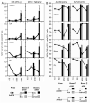
Figure 1. Sequential immunizations increase frequencies of antigen-specific T cells
(A) Schedule of immunization of BALB/c mice and time points of CD8+ T cell analyses. (B and C) Groups of 4 BALB/c mice were immunized with 1×10⁹ vp of indicated viruses encoding HIVgag. At different times post immunizations lymphocytes were isolated from spleen, popliteal and inguinal lymph nodes (LN), blood, peritoneal lavage (PL), mesenteric lymph nodes (MLN), Peyer's patches (PP) and small intestine and assayed for IFNγ production by ICS (B) or ELISpot (C) in response to gag peptide stimulation. The Xs connected by a line represent CD8+ T cell frequencies over time in mice that had a single immunization (either AdC68 or AdHu5), day 150 bars show frequencies after a single boost tested on day 150 after priming (filled bar: boosted with either AdHu5 or AdC68; gray bar: boosted with MVA), day 365 bars shows data after the second boost (filled bar: boosted with MVA; gray bar: boosted with AdHu5 in the AdC68 primed group or AdC68 in the AdHu5 primed group) tested on day 365 after priming. In all experiments shown in Figure 1 and subsequent Figures splenocytes from individual mice were analyzed and the graphs show either the average result, or in case of phenotypes (Fig 3A) a representative result from 4–5 samples. Lymphocytes from blood and the other tissues were pooled. For spleens, as noted in text when comparing values from a single immunization to boosts or from one boost to two boosts, p < 0.03.
Figure 2
Figure 2. An immunization regimen in which three heterologous vectors were given consecutively exhibits marked increase of antigen-specific T cells in gut mucosa
(A) Schedule of immunization of BALB/c mice and time points of CD8+ T cell analyses. (B) Groups of 3–5 BALB/c mice were immunized with 1×10⁹ vp AdC68HIVgag then boosted with the same dose of AdC7 and then AdHu5. All vectors encoded HIVgag. CD8+ T cells were isolated at indicated times from different tissues and stained with an antibody to CD8 and a HIVgag tetramer. The Xs connected by lines show gag-specific CD8+ T cell frequencies from mice over time after the 1st boost (tested on day 115 after priming), and the bars at d210 show frequencies in mice after the second boost (tested on day 210 after priming). For spleens, as noted in text when comparing values from a single immunization to boosts or from one boost to two boosts, p < 0.05.
Figure 3
Figure 3. History of antigen exposure does not result in significant changes in phenotypes of antigen-specific CD8+ T cells
(A and B) Lymphocytes isolated from spleen (SP), blood (BL) and draining lymph nodes (LN) of mice diagrammed in Figure 1A were stained with anti-CD8. They were also stained with antibodies to CD62L, CD27, or CD127 and HIVgag tetramer. (A) The data set represents mice that were primed with AdC68gag and then subsequently boosted with combinations of AdHu5 (H) and MVA (M) encoding HIVgag. The black line shows fluorescent intensity of tet+ CD8+ T cells and the shaded curve shows that of naïve CD8+ T cells. n.d. is not determined. (B) Frequencies of gag-specific CD8+ T cells from animals that were first immunized with AdC68gag or AdHu5gag. Data shown are for tet+ CD8+ cells. Xs connected by lines show frequencies over time in spleen of mice that had a single immunization, while bars at d150 show frequencies from spleens of mice that were boosted once and tested on day 150 after priming. The bars at d365 show frequencies after the second boost on day 365 after priming. (C) Phenotypes of lymphocytes isolated from mesenteric lymph nodes (MLN) and Peyer's patches (PP) from mice that received 3 immunizations as described in 2A. The black line shows fluorescent intensity of tet+ CD8+ T cells and the shaded curve shows that of naïve CD8+ T cells.
Figure 4
Figure 4. Multiple immunizations result in CD8+ T cells with increased functionality
(A) CD8+ T cells isolated from mice primed with AdC68gag. Mice described in Figure 1A were used to compare the percentage of CD8+ T cells that secreted IFNγ (left panel) and the percentage that were tet+ (right panel). Lymphocytes were isolated from spleen, draining lymph nodes (LN), blood (BL) and peritoneal lavage (PL). The Xs connected by lines show frequencies in mice after a single immunization with AdC68gag, bars at d150 represent frequencies after a single boost tested on day 150 after priming (filled bar is boosted with AdHu5 and shaded bar is boosted with MVA), bars at d365 represent data after a second boost tested on day 365 after priming (filled bar is boosted with MVA and shaded bar with AdHu5). (B) The same cells as (A) were assayed by ICS for percentage that secreted both IFNγ from both the AdC68gag and AdHu5gag primed groups. Gray bars indicate non AdHu5 pre-exposed animals and black bars indicate AdHu5 pre-exposed animals.
Figure 5
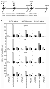
Figure 5
Effects of repeated immunization on the CD8+ T cell response in animals pre-exposed to viral vector. A group of BALB/c mice was pre-exposed to AdHu5 expressing rabies glycoprotein. Three weeks after exposure, titers to AdHu5 were measured and found to be between 1:80 and 1:360. Mice that were or were not pre-exposed to AdHu5 were then primed with 1×10⁹ vp of the indicated vectors expressing HIVgag, and 90 days post-prime lymphocytes were isolated from spleen, draining lymph nodes (LN), blood, and peritoneal lavage (PL) and tested for IFNγ production upon stimulation with a gag peptide by ICS. The remaining mice were boosted with vector. AdHu5 primed mice were boosted with AdC6, mice primed with AdC6 or AdC68 were boosted with AdHu5. On day 90 after the second boost, lymphocytes were isolated from boosted mice, as well as mice that did not receive a boost, were assayed for IFNγ production upon stimulation with a gag peptide by ICS. Shaded bars indicate non-AdHu5 pre-exposed mice and solid bars represent AdHu5 pre-exposed mice.

References

    1. Ahmed R, Gray D. Immunological memory and protective immunity: understanding their relation. Science. 1996;272(5258):54–60. - PubMed
    1. Badovinac VP, Porter BB, Harty JT. Programmed contraction of CD8(+) T cells after infection. Nat Immunol. 2002;3(7):619–26. - PubMed
    1. Casimiro DR, Bett AJ, Fu TM, Davies ME, Tang A, Wilson KA, Chen M, Long R, McKelvey T, Chastain M, Gurunathan S, Tartaglia J, Emini EA, Shiver J. Heterologous human immunodeficiency virus type 1 priming-boosting immunization strategies involving replication-defective adenovirus and poxvirus vaccine vectors. J Virol. 2004;78(20):11434–8. - PMC - PubMed
    1. Casimiro DR, Tang A, Perry HC, Long RS, Chen M, Heidecker GJ, Davies ME, Freed DC, Persaud NV, Dubey S, Smith JG, Havlir D, Richman D, Chastain MA, Simon AJ, Fu TM, Emini EA, Shiver JW. Vaccine-induced immune responses in rodents and nonhuman primates by use of a humanized human immunodeficiency virus type 1 pol gene. J Virol. 2002;76(1):185–94. - PMC - PubMed
    1. Cerwenka A, Morgan TM, Dutton RW. Naive, effector, and memory CD8 T cells in protection against pulmonary influenza virus infection: homing properties rather than initial frequencies are crucial. J Immunol. 1999;163(10):5535–43. - PubMed

Publication types

MeSH terms