

Effects of rapid eye movement sleep deprivation on hypocretin neurons in the hypothalamus of a rat model of depression
- PMID: 17590434
- PMCID: PMC2000483
- DOI: 10.1016/j.npep.2007.04.006
Effects of rapid eye movement sleep deprivation on hypocretin neurons in the hypothalamus of a rat model of depression
Abstract
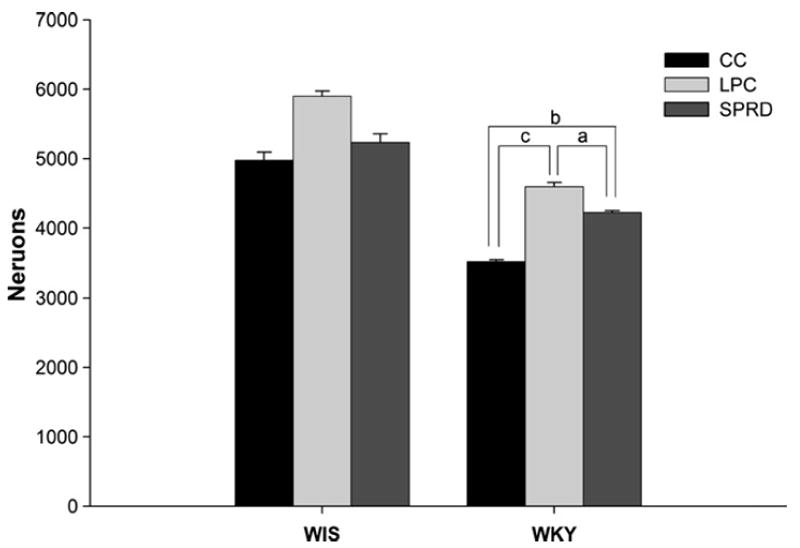
Hypocretin (Hcrt, also known as orexin) is a hypothalamic neuropeptide linked to narcolepsy, a disorder diagnosed by the appearance of rapid eye-movement sleep (REMS)-state characteristics during waking. Major targets of Hcrt-containing fibers include the locus coeruleus and the raphe nucleus, areas with important roles in regulation of mood and sleep. A relationship between REMS and mood is suggested by studies demonstrating that REMS-deprivation (REMSD) ameliorates depressive symptoms in humans. Additional support is found in animal studies where antidepressants and REMSD have similar effects on monoamiergic systems thought to be involved in major depression. Recently, we have reported that Wistar-Kyoto (WKY) rats, an animal model of depression, have reduced number and size of hypothalamic cells expressing Hcrt-immunoractivity compared to the parent, Wistar (WIS) strain, suggesting the possibility that the depressive-like attributes of the WKY rat may be determined by this relative reduction in Hcrt cells [Allard, J.S., Tizabi, Y., Shaffery, J.P., Trouth, C.O., Manaye, K., 2004. Stereological analysis of the hypothalamic hypocretin/orexin neurons in an animal model of depression. Neuropeptides 38, 311-315]. In this study, we sought to test the hypothesis that REMSD would result in a greater increase in the number and/or size of hypothalamic, Hcrt-immunoreactive (Hcrt-ir) neurons in WKY, compared to WIS rats. The effect of REMSD, using the multiple-small-platforms-over-water (SPRD) method, on size and number of Hcrt-ir cells were compared within and across strains of rats that experienced multiple-large-platforms-over-water (LPC) as well as to those in a normal, home-cage-control (CC) setting. In accord with previous findings, the number of Hcrt-ir cells was larger in all three WIS groups compared to the respective WKY groups. REMSD produced a 20% increase (p<0.02) in the number of hypothalamic Hcrt-ir neurons in WKY rats compared to cage control WKY (WKY-CC) animals. However, an unexpected higher increase in number of Hcrt-ir cells was also observed in the WKY-LPC group compared to both WKY-CC (31%, p<0.001) and WKY-SPRD (20%, p<0.002) rats. A similar, smaller, but non-significant, pattern of change was noted in WIS-LPC group. Overall the data indicate a differential response to environmental manipulations where WKY rats appear to be more reactive than WIS rats. Moreover, the findings do not support direct antidepressant-like activity for REMSD on hypothalamic Hcrt neurons in WKY rats.
Figures


References
-
- Adrien J, Dugovic C. Presence of a paradoxical sleep (PS) factor in the cerebrospinal fluid of PS-deprived rats. Eur J Pharmacol. 1984;100:223–226. - PubMed
-
- Allard JS, Tizabi Y, Shaffery JP, Trouth CO, Manaye K. Stereological analysis of the hypothalamic hypocretin/orexin neurons in an animal model of depression. Neuropeptides. 2004;38:311–315. - PubMed
-
- Anand A, Charney DS. Norepinephrine dysfunction in depression. J Clin Psychiatry. 2000;61(Suppl 10):16–24. - PubMed
-
- Basheer R, Magner M, McCarley RW, Shiromani PJ. REM sleep deprivation increases the levels of tyrosine hydroxylase and norepinephrine transporter mRNA in the locus coeruleus. Brain Res Mol Brain Res. 1998;57:235–240. - PubMed
-
- Benca RM. Mood disorders. In: Kryger MH, Roth T, Dement WC, editors. Principles and Practice of Sleep Medicine. Elsevier/Saunders; Philadelphia PA: 2005. pp. 1311–1326.
Publication types
MeSH terms
Substances
Grants and funding
LinkOut - more resources
Full Text Sources
Medical
