. 2007 Sep;27(17):5986-6000.
doi: 10.1128/MCB.00136-07. Epub 2007 Jun 25.

WAVE2 regulates high-affinity integrin binding by recruiting vinculin and talin to the immunological synapse

Affiliations

WAVE2 regulates high-affinity integrin binding by recruiting vinculin and talin to the immunological synapse

Jeffrey C Nolz et al. Mol Cell Biol. 2007 Sep.

Abstract

T-cell-receptor (TCR)-mediated integrin activation is required for T-cell-antigen-presenting cell conjugation and adhesion to extracellular matrix components. While it has been demonstrated that the actin cytoskeleton and its regulators play an essential role in this process, no mechanism has been established which directly links TCR-induced actin polymerization to the activation of integrins. Here, we demonstrate that TCR stimulation results in WAVE2-ARP2/3-dependent F-actin nucleation and the formation of a complex containing WAVE2, ARP2/3, vinculin, and talin. The verprolin-connecting-acidic (VCA) domain of WAVE2 mediates the formation of the ARP2/3-vinculin-talin signaling complex and talin recruitment to the immunological synapse (IS). Interestingly, although vinculin is not required for F-actin or integrin accumulation at the IS, it is required for the recruitment of talin. In addition, RNA interference of either WAVE2 or vinculin inhibits activation-dependent induction of high-affinity integrin binding to VCAM-1. Overall, these findings demonstrate a mechanism in which signals from the TCR produce WAVE2-ARP2/3-mediated de novo actin polymerization, leading to integrin clustering and high-affinity binding through the recruitment of vinculin and talin.


Figures
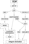
FIG. 1.
FIG. 1.
Signaling cascades required for TCR-mediated integrin activation. Ligation of the TCR activates the proximal tyrosine kinases Lck (not shown) and ZAP-70. These kinases can then activate several pathways known to regulate integrin activation, including the LAT-ITK-PLCγ1 pathway, required for both TCR-mediated calcium flux and activation of the small GTPase of the Ras family, RAP1. Targets of Rap1 (PKD, RAPL, MST1) regulate integrin activation primarily through the appropriate clustering of integrins on the cell surface. ADAP and SKAP55 are both required for TCR-mediated integrin activation and have been shown to affect both cytoskeletal dynamics as well as the localization of RAP1. ADAP and SKAP55 have also been shown to be indirectly linked to active RAP1 through another RAP1 effector, RIAM, which constitutively interacts with SKAP55. Finally, the RHO GTPases RAC1 and CDC42 (while never formally shown to be required for TCR-mediated integrin activation) can be activated by VAV1, leading to cytoskeletal reorganization, possibly through the activation of WAVE2. The integrin scaffolding protein Talin binds to F-actin filaments and also directly to the tail of the integrin β chain and has been shown to control both integrin affinity and clustering. Importantly, several of these proteins (*) are not required for PMA-stimulated integrin activation, suggesting that integrins can be activated independently of proximal TCR-stimulated signaling pathways when stimulated with PMA. Whether WAVE2 regulates cytoskeletal rearrangement leading to integrin activation is the focus of this study.
FIG. 2.
FIG. 2.
WAVE2 is not required for TCR-mediated activation of VAV1, RAC1, or CDC42. (A) Jurkat T cells were transfected with either control or WAVE2-suppression vector. At 72 h posttransfection, cells were stimulated with anti-CD3/goat anti-mouse antibody for 0, 1, 2, 5, or 15 min. Cells were immediately lysed, and after clarification, VAV1 was immunoprecipitated (IP). Bound proteins were eluted, separated by SDS-PAGE, transferred, and blotted with 4G10 MAb (pTyr) and VAV1. Whole-cell extracts (WCE) were also analyzed for expression of WAVE2 and VAV1. (B and C) Cells were transfected as described for panel A. Cells were stimulated for 0, 1, or 5 min and immediately lysed. Following clarification, GTP-bound RAC1 (B) or GTP-bound CDC42 (C) was detected using glutathione S-transferase-PAK GTPase binding domain and immunoblotting.
FIG. 3.
FIG. 3.
The VCA domain of WAVE2 regulates TCR-mediated integrin activation. (A) WAVE2 contains an N-terminal WAVE-homology domain, followed by a small BR, a large PR, and a C-terminal VCA domain. Jurkat T cells were transfected with “suppression/reexpression” constructs which suppress endogenous levels of WAVE2 and reexpress FLAG-tagged shRNA-resistant forms of WAVE2 and various deletion mutants. At 72 h posttransfection, whole-cell extracts were harvested, separated by SDS-PAGE, transferred, and blotted for WAVE2, FLAG, and ARPC2 as a loading control. The immunoreactive portion of WAVE2 is not present in the ΔPR mutant and can only be detected with FLAG immunoblotting. Multiple bands in the ΔVCA and ΔBR versions of WAVE2 are most likely due to different phosphorylation states of the proteins (36). (B) Cells were transfected as described for panel A and analyzed for their ability to adhere to fibronectin in response to stimulation of the TCR (+) or in the absence of stimulation (−). Adherent cells (GFP negative, GFP low, and GFP high) were detected using flow cytometry. GFP-negative cells (untransfected) served as an internal control to demonstrate adequate stimulation required for adhesion. (C) Cells were transfected as described for panel A and analyzed for their ability to form conjugates with PKH26-stained Raji B cells that were either pulsed with SEE or left unpulsed. Conjugate formation was determined using two-color flow cytometry as described in Materials and Methods. (D) Cells were transfected as described for panel A with either control or WAVE2-suppression vector and analyzed for surface expression of both β1 and β2 integrins. Cells were gated as GFP positive or GFP negative and stained with control IgG (white histogram), β1 (black histogram), or β2 (gray histogram) integrin in the different cell populations.
FIG. 4.
FIG. 4.
The VCA domain of WAVE2 regulates actin polymerization and integrin activation downstream of proximal signals, but this occurs independently of calcium mobilization. (A) Jurkat T cells were transfected with control, WAVE2-suppression, WT WAVE2-reexpression, or ΔVCA WAVE2-reexpression vectors and allowed to recover for 72 h. T cells (green) were then allowed to form conjugates with CMAC-stained Raji B cells (blue) that were pulsed with SEE, allowed to adhere to poly-l-lysine-coated coverslips, fixed, and stained with rhodamine phalloidin (red) to visualize the accumulation of F-actin at the IS. Accumulation of F-actin was quantified in 50 random conjugates as described in Materials and Methods; data represent the average results of three independent experiments. (B) Jurkat T cells were transfected as described for panel A. After 72 h, calcium measurements were performed using single-cell fluorescence ratio (Fura-2 imaging) of GFP-positive control and WAVE2-suppressed Jurkat cells. In a calcium-free bath solution, increases in the Fura-2 ratio reflect Ca2+ release from intracellular stores. Extracellular Ca2+ entry via activated calcium release-activated calcium channels was subsequently assessed by reintroduction of extracellular calcium (2 mM Ca2+). Data represent the results of three independent experiments for each condition, and each trace represents the average response of at least 100 cells in the recording chamber. (C) Cells were transfected as described for panel A and analyzed for their ability to adhere to fibronectin in response to stimulation with 10 ng PMA/ml. Adhesion was determined as described for Fig. 2B.
FIG. 5.
FIG. 5.
Localization of both LFA-1 and β1 integrins to the IS requires WAVE2 and ARP2/3. (A) Jurkat T cells were transfected with control, WAVE2 suppression, or ARP2 suppression vectors and allowed to recover for 72 h. T cells (green) were then allowed to form conjugates with CMAC-stained Raji B cells (blue) that were pulsed with SEE, allowed to adhere to poly-l-lysine-coated coverslips, fixed, and stained with antibody against the β2 chain of LFA-1. Localization of LFA-1 to the synapse was quantified using 50 random conjugates as described in Materials and Methods; data represent the average results of three independent experiments. (B) Cells were transfected and conjugates were formed as described for panel A. Conjugates were then analyzed for their ability to localize β1 integrins to the IS, and quantification was determined as described for panel A.
FIG. 6.
FIG. 6.
The ARP2/3 complex is required to link WAVE2 to vinculin. (A) Human CD4+ T cells were allowed to form conjugates with CMAC-labeled NALM6 B cells (blue) that were pulsed with a superantigen cocktail containing SEE, SEA, SEB, and SEC (+ SAg) or left unpulsed (− SAg). Conjugates were then allowed to adhere to poly-l-lysine-coated coverslips, fixed, and subsequently stained for vinculin (red) and F-actin (green) or (B) vinculin (red) and WAVE2 (green). (C) Jurkat T cells were transfected with either control vector or ARP2-suppression vector and allowed to recover for 72 h. Cells were then left unstimulated or were stimulated with anti-CD3/CD28 for 30 min and immediately lysed. Lysates were clarified, and vinculin was immunoprecipitated (IP). Bound proteins were eluted, separated by SDS-PAGE, transferred, and subsequently blotted for expression of WAVE2, ARP2, and vinculin. Whole-cell extracts (WCE) were also analyzed for protein levels of WAVE2, ARP2, and vinculin. (D) Cells were transfected with control, WAVE2 suppression, WT WAVE2 reexpression, or ΔVCA WAVE2 reexpression vector. Cells were stimulated, and lysates were immunoprecipitated as described for panel C and subsequently blotted for WAVE2, ARPC2, and vinculin.
FIG. 7.
FIG. 7.
Vinculin is required for TCR-mediated integrin activation occurring independently of actin and integrin accumulation at the IS. (A) Jurkat T cells were transfected with either vector control or with suppression vectors against vinculin. At 72 h posttransfection, cells were analyzed by immunoblotting for expression of vinculin and WAVE2. (B) Cells were transfected as described for panel A and analyzed for their ability to form conjugates with PKH26-stained Raji B cells that were either pulsed with SEE or left unpulsed. Conjugate formation was determined using two-color flow cytometry as described in Materials and Methods. (C) Jurkat T cells were transfected as described for panel A. T cells (green) were then allowed to form conjugates with SEE-pulsed Raji B cells (blue), allowed to adhere to poly-l-lysine-coated coverslips, fixed, and stained with rhodamine phalloidin to visualize accumulation of F-actin at the IS. Actin polymerization was quantified using 50 random conjugates as described in Materials and Methods; data represent the average results of two independent experiments. (D) Same as panel C except that the cells were stained for β2 integrin.
FIG. 8.
FIG. 8.
The association between ARP2/3 and vinculin is required for TCR-mediated integrin activation. (A) Jurkat T cells were transfected with control, vinculin suppression, WT vinculin reexpression, or P878A vinculin reexpression vectors. At 72 h posttransfection, whole-cell extracts were harvested, separated by SDS-PAGE, and transferred, and membranes were subsequently blotted for vinculin, FLAG, and ARPC2 as a loading control. (B) Cells were transfected as described for panel A and were subjected to TCR stimulation (+) or were left unstimulated (−) and were analyzed for their ability to adhere to fibronectin. (C) Cells were transfected as described for panel A and were subjected to stimulation with 10 ng/ml PMA or were left unstimulated and were analyzed for their ability to adhere to fibronectin. Adherent cells were detected using flow cytometry as described for Fig. 3B.
FIG. 9.
FIG. 9.
Vinculin associates with WAVE2, ARP2/3, and talin in response to TCR stimulation. (A) Jurkat T cells were stimulated with anti-CD3/CD28 antibody for 0, 5, 15, or 30 min and lysed, and immunoprecipitations (IP) were performed using either an isotype control IgG antibody or an antivinculin antibody. Bound proteins were eluted, separated by SDS-PAGE, and transferred, and membranes were subsequently blotted for talin, WAVE2, ARPC2, and vinculin. (B) Same as panel A except that human CD4+ T cells were used. (C) Jurkat T cells were transfected with either control or WAVE2 suppression vectors and allowed to recover for 72 h. Cells were then stimulated with anti-CD3/CD28 for 0, 15, or 30 min and lysed, and β1 integrin was immunoprecipitated. Bound proteins were eluted, separated by SDS-PAGE, and transferred, and membranes were subsequently blotted for talin, vinculin, and β1 integrin. Whole-cell extracts (WCE) were also analyzed for expression of talin, vinculin, β1 integrin, and WAVE2.
FIG. 10.
FIG. 10.
The VCA domain of WAVE2 and vinculin are required for talin localization and activation-dependent high-affinity integrin binding. (A) Jurkat T cells were transfected with control, WAVE2 suppression, WT WAVE2 reexpression, or ΔVCA WAVE2 reexpression vectors and allowed to recover for 72 h. T cells (green) were then allowed to form conjugates with CMAC-stained Raji B cells (blue) that were pulsed with SEE, allowed to adhere to poly-l-lysine coated coverslips, fixed, and stained with antibody against talin (red) to visualize accumulation at the IS. Localization of talin was quantified using 50 random conjugates as described in Materials and Methods; data represent the average results of three independent experiments. (B) Same as panel A except that cells were transfected with control or vinculin suppression vectors. Localization was quantified as described for panel A. (C) Jurkat T cells were transfected with control, WAVE2 suppression, or vinculin suppression vectors and allowed to recover for 72 h. Cells were then either left unstimulated or stimulated with 50 ng/ml PMA or 1 mM Mn2+ for 10 min in the presence of soluble VCAM-1-Fc. Cells were then diluted and immediately fixed with paraformaldehyde and subsequently stained to detect binding of soluble VCAM-1 as described in Materials and Methods. Cells were analyzed by flow cytometry to detect binding of VCAM-1 and separated based on GFP expression levels. Gated populations indicate cells that bind with either high (right) or low (left) affinity to VCAM-1. High, Low, and Neg (negative) (listed to the left of each stimulation condition panel) refer to the GFP-expressing populations being analyzed in the experiment.
FIG. 11.
FIG. 11.
Proposed model for the role of WAVE2, ARP2/3, vinculin, talin, and actin polymerization in TCR-mediated integrin activation. (A) In resting cells, WAVE2, ARP2/3, vinculin, and talin are free of association and cell surface integrins are distributed evenly on the cell surface. (B) Stimulation of the TCR causes the activation of several signaling cascades leading to the activation of WAVE2, which then binds ARP2/3 and promotes de novo actin polymerization for the forming IS. Vinculin localizes to the IS through a direct interaction with WAVE2 and the ARP2/3 complex, and integrins begin to localize to the pSMAC. (C) Finally, talin (probably through its association with vinculin and F-actin) localizes and binds both β1 and β2 integrins, resulting in the high-affinity conformation at the IS. The newly formed F-actin generated by WAVE2 probably also serves as a binding partner for both vinculin and talin, providing the structural integrity required for stabilizing the integrin at the pSMAC.

References

    1. Calderwood, D. A., R. Zent, R. Grant, D. J. Rees, R. O. Hynes, and M. H. Ginsberg. 1999. The Talin head domain binds to integrin beta subunit cytoplasmic tails and regulates integrin activation. J. Biol. Chem. 274:28071-28074. - PubMed
    1. Campbell, I. D., and M. H. Ginsberg. 2004. The talin-tail interaction places integrin activation on FERM ground. Trends Biochem. Sci. 29:429-435. - PubMed
    1. Cannon, J. L., and J. K. Burkhardt. 2004. Differential roles for Wiskott-Aldrich syndrome protein in immune synapse formation and IL-2 production. J. Immunol. 173:1658-1662. - PubMed
    1. Cannon, J. L., C. M. Labno, G. Bosco, A. Seth, M. H. McGavin, K. A. Siminovitch, M. K. Rosen, and J. K. Burkhardt. 2001. Wasp recruitment to the T cell:APC contact site occurs independently of Cdc42 activation. Immunity 15:249-259. - PubMed
    1. Cao, Y., E. M. Janssen, A. W. Duncan, A. Altman, D. D. Billadeau, and R. T. Abraham. 2002. Pleiotropic defects in TCR signaling in a Vav-1-null Jurkat T-cell line. EMBO J. 21:4809-4819. - PMC - PubMed

Publication types

MeSH terms

LinkOut - more resources