. 2007 Aug 31;282(35):25779-89.
doi: 10.1074/jbc.M702277200. Epub 2007 Jun 25.

Exosomes released from infected macrophages contain Mycobacterium avium glycopeptidolipids and are proinflammatory

Affiliations

Exosomes released from infected macrophages contain Mycobacterium avium glycopeptidolipids and are proinflammatory

Sanchita Bhatnagar et al. J Biol Chem. .

Abstract

Mycobacterium avium is a major opportunistic pathogen in HIV-positive individuals and is responsible for increased morbidity and mortality in AIDS patients. M. avium express glycopeptidolipids (GPLs) as a major cell wall constituent, and recent studies suggest that GPLs play an important role in M. avium pathogenesis. In the present study we show that M. avium-infected macrophages release GPLs, which are trafficked from the phagosome through the endocytic network to multivesicular bodies. Prior studies have shown that multivesicular bodies can fuse with the plasma membrane releasing small 50 to 100 nm vesicles known as exosomes. We found that M. avium-infected macrophages release exosomes containing GPLs leading to the transfer of GPLs from infected to uninfected macrophages. Interestingly, exosomes isolated from M. avium-infected but not from uninfected macrophages can stimulate a proinflammatory response in resting macrophages. This proinflammatory response is dependent on Toll like receptor (TLR) 2, TLR4, and MyD88 suggesting that released exosomes contain M. avium-expressed TLR ligands. Our studies are the first to demonstrate that exosomes isolated from mycobacteria-infected macrophages can induce a proinflammatory response, and we hypothesize that exosomes play an important role in immune surveillance during intracellular bacteria infections.


Figures
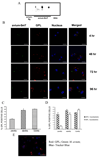
FIGURE 1
FIGURE 1. Intracellular trafficking of glycopeptidolipids in macrophages infected with M. avium serovar 2
A, purified GPLs extracted from three separate Smooth-Transparent M. avium 2151 (avium-SmT) colonies were separated by TLC and probed with serovar 2-specific anti-GPL. A similar extraction protocol was followed using the GPL-deficient Rough isolate of M. avium 2151 (avium-Rg). Bovine serum albumin was used as a control. B, BMMs were infectedwithFITC-labeledSmTM.avium 2151for4h,washed,andincubatewithfreshmediaforthetimesindicated. Fixed cells were stained for GPL using the serovar 2-specific anti-GPL and visualized by confocal microscopy. Nuclei were stained using the bisalkylaminoanthraquinone fluorphore Draq5. Coincident staining appears yellow in the merged images. C, quantitation of the BMMs that stained positive for GPL or D, BMMs that stained positive for GPL alone or GPL andM. avium.Aminimal of 150 cells were counted for each sample and expressed as mean±S.D. from three separate experiments. E,BMMs infected with FITC-labeled M. avium 2151 were incubated with equal number of uninfected macrophages, labeled with cell tracker blue 7-amino-4-chloromethylcoumarin. Cells were stained for GPL as described above. Data are representative of three separate experiments (scale = 10µm).
FIGURE 2
FIGURE 2. GPLs localize to multivesicular bodies in SmT M. avium 2151-infected macrophages
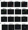
A, cells infected for 48 h with SmT M. avium 2151 were fixed, permeabilized, and probed using antibodies against LAMP-1,MHCclass II, GPL, and Grp78. B,BMMs were infected with M. avium 2151 for 48 h and pulsed for 4 h with N-Rh-PE. Infected and stained cells were visualized by confocal microscopy. Nuclei were labeled with Draq5. Coincident staining appears yellow in the merged images. Images are representative of three separate experiments (scale = 10 µm).
FIGURE 3
FIGURE 3. Rab11 transiently colocalizes with GPL-containing vesicles
Cells were permeabilized and fixed 24, 48, or 72 h post-infection with M. avium 2151. Cells were probed with anti-Rab11 and anti-GPL antibody. Antibody-stained cells were visualized by confocal microscopy. Nuclei were labeled with Draq5. Coincident staining appears yellow in the merged images. Images are representative of three separate experiments (scale = 10 µm).
FIGURE 4
FIGURE 4. Extracellular release of GPL-containing exosomes
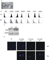
A, transmission electron micrographs of purified exosomes isolated from uninfected J774 cells. Scale bar: 100 nm. B, exosomes isolated from the culture supernatants of SmT M. avium 2151-infected (IN) or uninfected (UI) BMM were coated onto sulfate/aldehyde latex beads and stained with FITC-conjugated antibodies specific for the indicated protein (closed peaks) or the corresponding isotype-matched control antibody (open peaks). Beads were analyzed by flow cytometry, and mean fluorescent intensities are shown. Data is representative of four separate experiments. C, 10 µg of exosomes were analyzed by Western blot for the presence of the mycobacteria LAM and the host protein hsp70. D, 10µg of purified exosomes isolated from uninfected or serovar 2 M. avium 2151, serovar 1 M. avium 101, or serovar 4 M. avium A5-infected macrophages were added to BALB/c BMMs. 24 h post-treatment, cells were fixed, permeabilized, and labeled with serovar-specific anti-GPL antibody. Nuclei were labeled with Draq5. Fluorescent images are representative of two separate experiments. SmT, M. avium 2151 SmT strain (scale = 10 µm).
FIGURE 5
FIGURE 5. Exosomes from M. avium 2151-infected macrophages induce a MyD88 dependent pro-inflammatory response in primary macrophages
Exosomes isolated from M. avium 2151-infected or uninfected J774 cells were added to BALB/c BMMs and 24 h post-treatment culture supernatants were assayed for TNF-α (A) or RANTES (B) by ELISA. Data are representative of five separate experiments. C, WT and MyD88−/− C57BL/6 BMMs were treated with 1 µg or 5 µg of exosome isolated from uninfected or SmT M. avium 2151-infected macrophages. TNF-α levels were measured 24 h post-exosome treatment by ELISA. Data are representative of three separate experiments, and ELISA values are expressed as mean ± S.D. for duplicate wells. *, significant to exosomes from uninfected cells (p < 0.01).
FIGURE 6
FIGURE 6. Exosomes and not apoptotic vesicles are responsible for the proinflammatory activity
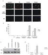
A, J774 cells were infected with M. avium for 4 h. As controls macrophages were left uninfected or were starved of fetal calf serum to induce apoptosis. Macrophages were fixed and stained for annexin V (green) and propidium iodide (red). Nuclei were stained using Draq5. Fluorescence images are representative of two independent experiments (bar = 10µm). B, exosomes were prepared from J774 cells infected with M. avium and treated with 50µm of the caspase-3 inhibitor Ac-DEVD-CHO or left untreated. As a control, apoptotic vesicles were also purified from serum-deprived J774 cells. Exosomes or apoptotic vesicles were added to separate, uninfected J774 cells, and 24 h post-treatment, culture supernatants were assayed for TNF-α by ELISA. Data are representative of two independent experiments and are expressed as mean±S.D. for duplicate wells. C, cell lysates obtained 24 h post-treatment with exosomes or apoptotic vesicles were analyzed for iNOS expression by Western blot. Total p38 was used as a loading control. Results are representative of two separate experiments. D, sulfate/aldehyde latex beads coated with exosomes or apoptotic vesicles were probed by flow cytometry for annexin V binding or FcγRII/III and the percentage of beads positive for the markers shown. Results are representative of two independent experiments. UI, uninfected J774 cells; SmT, J774 cells infected with SmT M. avium 104.
FIGURE 7
FIGURE 7. The presence of GPLs on exosomes is not required for exosome-induced TNF-α production
BALB/c BMMs were incubated with exosomes isolated from macrophages infected with SmT (GPL-sufficient) or Rg (GPL-deficient) M. avium 2151. TNF-α production by the infected BMMs were measured 24 h post treatment by ELISA. Data are representative of three separate experiments, and ELISA values are expressed as mean ± S.D. for duplicate wells. *, significant to exosomes from uninfected cells (p < 0.01).

References

    1. Horsburgh CR., Jr J. Infect. Dis. 1999;179(Suppl 3):S461–S465. - PubMed
    1. Rusin PA, Rose JB, Haas CN, Gerba CP. Rev. Environ. Contam. Toxicol. 1997;152:57–83. - PubMed
    1. Frehel C, de Chastellier C, Lang T, Rastogi N. Infect. Immun. 1986;52:252–262. - PMC - PubMed
    1. Fratti RA, Vergne I, Chua J, Skidmore J, Deretic V. Electrophoresis. 2000;21:3378–3385. - PubMed
    1. Fratti RA, Chua J, Vergne I, Deretic V. Proc. Natl. Acad. Sci. U. S. A. 2003;100:5437–5442. - PMC - PubMed

Publication types

MeSH terms