. 2007 Jul;48(7):3107-14.
doi: 10.1167/iovs.06-0955.

Horizontal saccade disconjugacy in strabismic monkeys

Affiliations

Horizontal saccade disconjugacy in strabismic monkeys

LaiNgor Fu et al. Invest Ophthalmol Vis Sci. 2007 Jul.

Abstract

Purpose: Previous studies have shown that binocular coordination during saccadic eye movement is affected in humans with large strabismus. The purpose of this study was to examine the conjugacy of saccadic eye movements in monkeys with sensory strabismus.

Methods: The authors recorded binocular eye movements in four strabismic monkeys and one unaffected monkey. Strabismus was induced by first occluding one eye for 24 hours, switching the occluder to the fellow eye for the next 24 hours, and repeating this pattern of daily alternating monocular occlusion for the first 4 to 6 months of life. Horizontal saccades were measured during monocular viewing when the animals were 2 to 3 years of age.

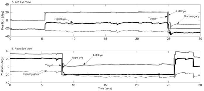
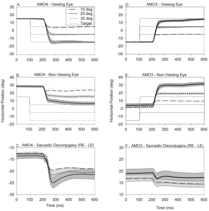
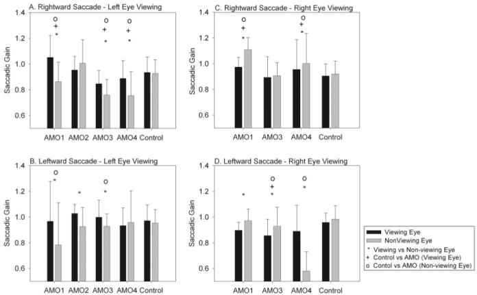
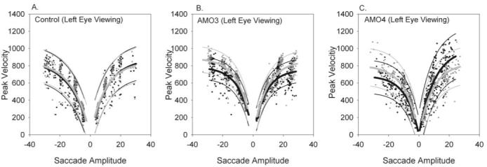
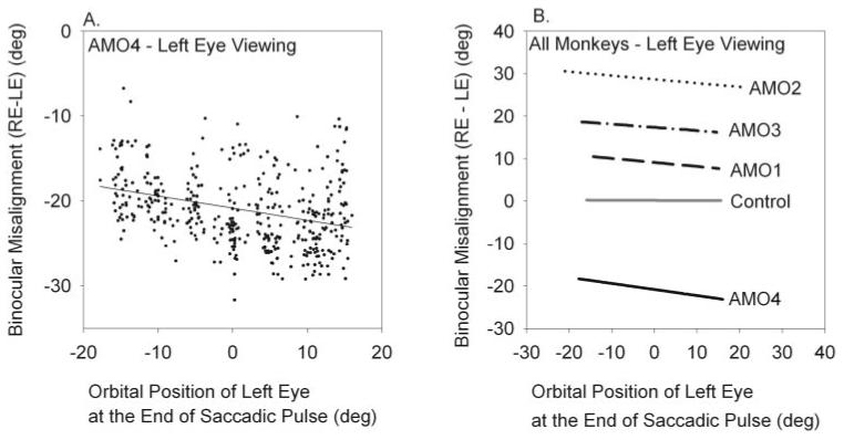
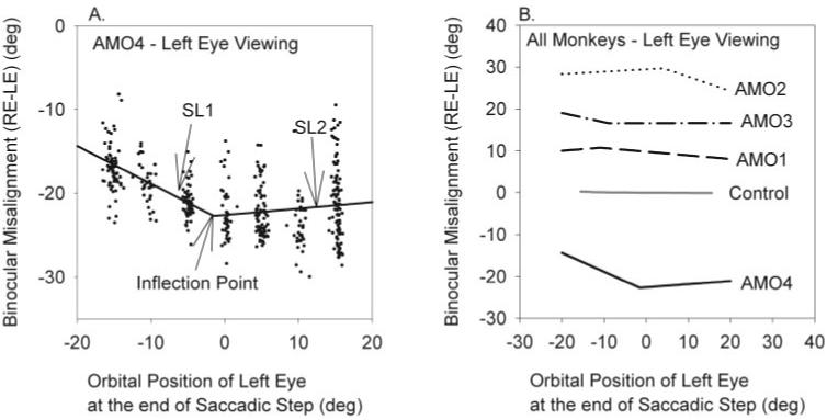
Results: Horizontal saccade testing during monocular viewing showed that the amplitude of saccades in the nonviewing eye was usually different from that in the viewing eye (saccade disconjugacy). The amount of saccade disconjugacy varied among animals as a function of the degree of ocular misalignment as measured in primary gaze. Saccade disconjugacy also increased with eccentric orbital positions of the nonviewing eye. If the saccade disconjugacy was large, there was an immediate postsaccadic drift for less than 200 ms. The control animal showed none of these effects.

Conclusions: As do humans with large strabismus, strabismic monkey display disconjugate saccadic eye movements. Saccade disconjugacy varies with orbital position and increases as a function of ocular misalignment as measured in primary gaze. This type of sensory-induced strabismus serves as a useful animal model to investigate the neural or mechanical factors responsible for saccade disconjugacy observed in humans with strabismus.


Figures

Figure 1
Figure 1
Eye position records for esotropic monkey AMO4 during monocular viewing with the left eye (A) and with the right eye (B). Traces indicate target position (black light line), right-eye position (black heavy line), left-eye position (gray heavy line), and disconjugacy (gray light line). Disconjugacy or ocular misalignment was determined by subtracting right-eye position from left-eye position. Positive values indicate rightward positions, and negative values indicate leftward positions. The figure illustrates that saccade are disconjugate and that ocular misalignment appears to vary with orbital position.
Figure 2
Figure 2
(A-C) Saccadic eye movement trajectories made to three different leftward target steps (10°, 20°, and 30°) from the same orbital eye position in esotropic monkey AMO4 when the right eye was viewing (left eye was patched). During straight-ahead binocular viewing, this animal had esotropia of 21°. Positive values indicate rightward eye position, and negative values indicate leftward eye position. (A) Trajectory of the viewing (right) eye. (B) Trajectory of the nonviewing (left) eye. (C) Degree of ocular misalignment (right-eye position - left-eye position). (D-F) Similar data from exotropic monkey AMO3 for rightward target steps during left-eye viewing. Both animals displayed disconjugate saccadic eye movements and postsaccadic drift that was orbital position dependent. Therefore, ocular misalignment before the saccade, at the end of the saccade pulse (i.e., after initial saccade), and at the end of the saccadic step (i.e., after postsaccadic drift) are all different. In all panels, trials are aligned on saccade onset.
Figure 3
Figure 3
(A, B) Rightward and leftward saccadic pulse gains in all monkeys for the viewing eye and the nonviewing eye for left-eye viewing. (C, D) Rightward and leftward saccadic pulse gains in all monkeys for the viewing eye and the nonviewing eye for right-eye viewing condition. Statistically significant differences (P < 0.05) obtained from multiple comparisons (○, +, *). The main finding is that there were differences in the pulse gain between viewing and nonviewing eyes in the strabismic animals, though the differences were idiosyncratic.
Figure 4
Figure 4
Amplitude-peak velocity relationship in viewing (gray triangles) and nonviewing (black circles) eyes in control animal (A), exotropic animal AMO3 (B), and esotropic animal AMO4 (C). Data for these figures were obtained during monocular left-eye viewing. Also plotted are exponential curve fits (heavy lines), along with 95% prediction bands (light lines). The gray lines are fits to the viewing eye data, and black lines are fits to the nonviewing eye data. There is significant overlap in the prediction intervals for the viewing and nonviewing eyes in the control animal, esotrope, and exotrope, suggesting that there was no difference in amplitude-peak velocity main-sequence relationship between viewing and nonviewing eyes. The main-sequence relationship was also similar when the strabismic animals were compared with the control animal.
Figure 5
Figure 5
(A) Relation of binocular misalignment immediately at the end of the saccadic pulse (disconjugacy of the saccadic pulse) with orbital position in AMO4 during left-eye viewing. Black dots indicate each saccade. The black line represents linear regression to the saccade data. (B) Summary of fits for all our monkeys obtained during the left-eye viewing condition showing a linear relationship between disconjugacy of saccadic pulse and orbital position. The orbital position of the viewing (left) eye at the end of initial saccade is plotted on the x-axis. The amount of misalignment at the end of initial saccade is plotted on the y-axis. Details about each of the fits such as slopes and goodness-of-fit are provided in Table 1.
Figure 6
Figure 6
Relation of binocular misalignment at the end of the saccadic step (i.e., after postsaccadic drift) with orbital position. (A) Data collected in AMO4 during left-eye viewing. Black dots indicate each saccade. Because the data indicated that ocular misalignment was comitant for certain orbital positions after the saccadic step, we used two-segment, piecewise linear regression to fit all the saccade data. That portion of the line that varied with orbital eye position was labeled SL1. That portion of the line that varied very little with orbital eye position was labeled SL2. The inflection point is the orbital position at which the slopes changed, as determined by regression analysis. (B) Summary of two-segment fits of saccade data after saccadic step obtained during left-eye viewing. In each of the AMO animals, we were able to identify a region of relative comitancy. Details about each of the fits, such as slopes SL1, SL2, inflection point, and the goodness-of-fit are provided in Table 1.

References

    1. von Noorden GK. Binocular Vision and Ocular Motility: Theory and Management of Strabismus. 5th ed. CV Mosby; St. Louis: 1996.
    1. Leigh RJ, Zee DS. The Neurology of Eye Movements. 3rd ed. Oxford University Press; New York: 1999. Contemporary Neurology Series.
    1. Boothe RG, Dobson V, Teller DY. Postnatal development of vision in human and nonhuman primates. Ann Rev Neurosci. 1985;8:495–545. - PubMed
    1. Lorenz B. Genetics of isolated and syndromic strabismus: facts and perspectives. Strabismus. 2002;10:147–156. - PubMed
    1. Maxwell GF, Lemij HG, Collewijn H. Conjugacy of saccades in deep amblyopia. Invest Ophthalmol Vis Sci. 1995;36:2514–2522. - PubMed

Publication types