

Presence of an established calcification marker in trabecular meshwork tissue of glaucoma donors
- PMID: 17591888
- PMCID: PMC1994153
- DOI: 10.1167/iovs.06-1403
Presence of an established calcification marker in trabecular meshwork tissue of glaucoma donors
Abstract
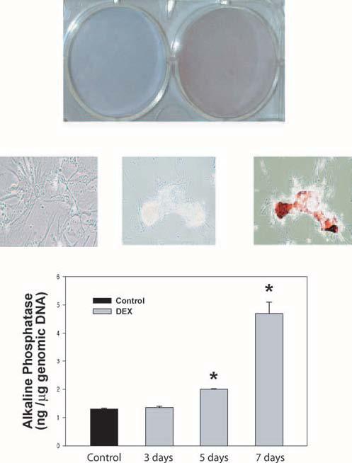
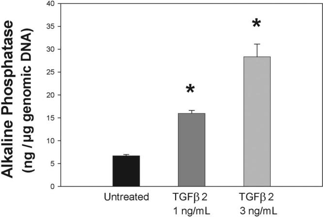
Purpose: To determine the presence of calcification markers in the trabecular meshwork tissue from glaucoma donors and in trabecular meshwork cells insulted by dexamethasone (DEX) and transforming growth factor beta2 (TGFbeta2), factors associated with glaucoma. To investigate as well the effect of silencing the inhibitor of calcification matrix Gla (MGP) in the trabecular meshwork cells.
Methods: Trabecular meshwork tissue was obtained from perfused postmortem anterior segments of glaucomatous and normal eyes. Primary trabecular meshwork cells were obtained from residual corneal rims after surgical corneal transplantation. Calcification marker alkaline phosphatase (ALP) enzyme activity was assayed by fluorescence produced after substrate cleavage. DNA quantification was evaluated by fluorescence produced after binding to the Hoechst dye. Transfection of siRNA to primary cells was accomplished by nucleofector electroporation with trabecular meshwork-optimized conditions. cDNA quantification was performed with the use of TaqMan real-time PCR.
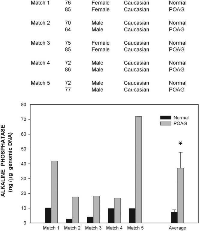
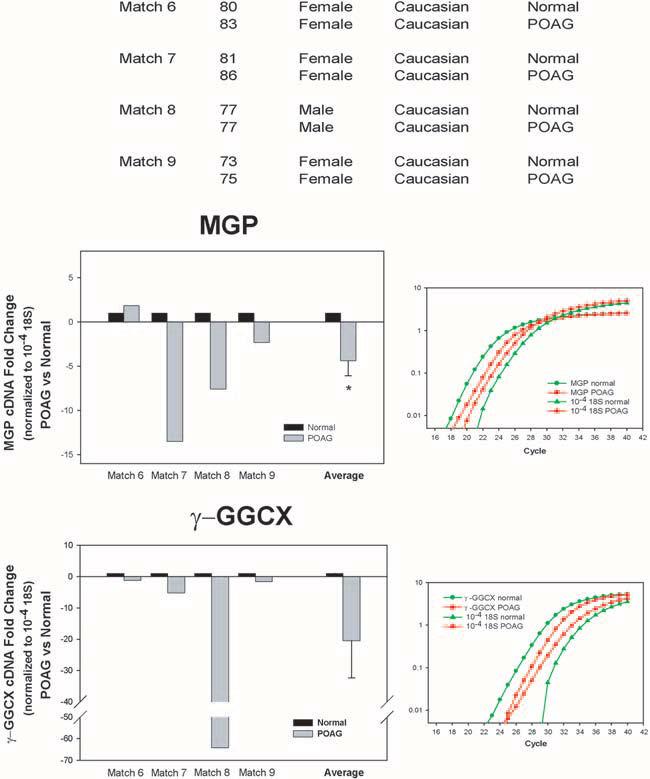
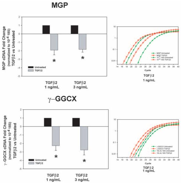
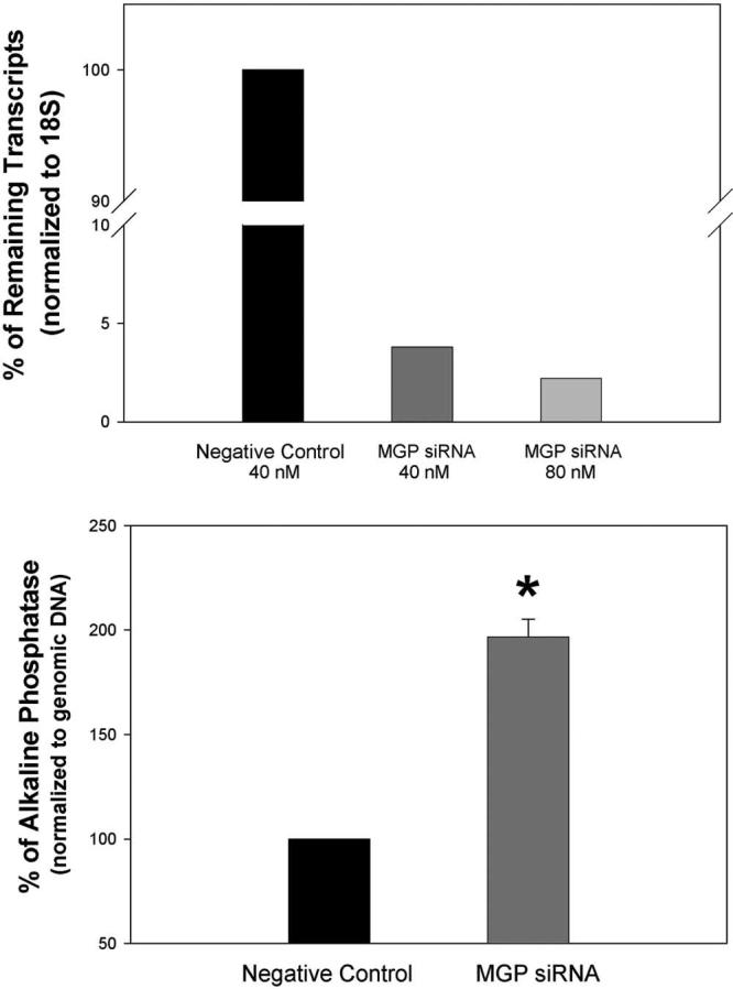
Results: Human trabecular meshworks from glaucoma donors exhibited significantly higher levels of ALP activity than their matched counterparts with normal eyes. The normalized ALP of the control specimens was 7.3 +/- 1.6 ng ALP/microg DNA (n = 4), whereas that of the glaucomatous tissue was 37.0 +/- 10.7 ng ALP/microg genomic DNA (n = 5; P </= 0.04). DEX and TGFbeta2 significantly induced the upregulation of ALP activity in two trabecular meshwork primary cell lines. Expression of the gene encoding MGP was reduced in the glaucomatous tissue by -4.4 +/- 1.7-fold (n = 9; P </= 0.006). Silencing MGP by siRNA resulted in ALP activity that was increased by 197% +/- 8.4% (P </= 0.0003).
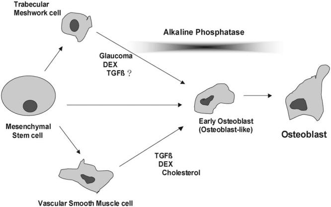
Conclusions: The increased activity of the calcification marker, ALP, in glaucomatous trabecular meshworks might be indicative of an undergoing mineralization process during development of the disease. Inhibition of the calcification mechanism represented by the presence of active MGP appears to be compromised in glaucomatous tissue.
Figures
Similar articles
-
Functional delivery of synthetic naked siRNA to the human trabecular meshwork in perfused organ cultures.Mol Vis. 2007 Aug 1;13:1363-74. Mol Vis. 2007. PMID: 17768383
-
Matrix GLA protein function in human trabecular meshwork cells: inhibition of BMP2-induced calcification process.Invest Ophthalmol Vis Sci. 2006 Mar;47(3):997-1007. doi: 10.1167/iovs.05-1106. Invest Ophthalmol Vis Sci. 2006. PMID: 16505034 Free PMC article.
-
Transforming growth factor-beta2 utilizes the canonical Smad-signaling pathway to regulate tissue transglutaminase expression in human trabecular meshwork cells.Exp Eye Res. 2011 Oct;93(4):442-51. doi: 10.1016/j.exer.2011.06.011. Epub 2011 Jun 24. Exp Eye Res. 2011. PMID: 21722634 Free PMC article.
-
A single gene connects stiffness in glaucoma and the vascular system.Exp Eye Res. 2017 May;158:13-22. doi: 10.1016/j.exer.2016.08.022. Epub 2016 Sep 1. Exp Eye Res. 2017. PMID: 27593913 Free PMC article. Review.
-
Evidence for a calcification process in the trabecular meshwork.Exp Eye Res. 2009 Apr;88(4):738-46. doi: 10.1016/j.exer.2008.11.027. Epub 2008 Dec 6. Exp Eye Res. 2009. PMID: 19084518 Free PMC article. Review.
Cited by
-
Development of a model of elevated intraocular pressure in rats by gene transfer of bone morphogenetic protein 2.Invest Ophthalmol Vis Sci. 2013 Aug 13;54(8):5441-55. doi: 10.1167/iovs.13-11651. Invest Ophthalmol Vis Sci. 2013. PMID: 23821199 Free PMC article.
-
Consensus recommendations for trabecular meshwork cell isolation, characterization and culture.Exp Eye Res. 2018 Jun;171:164-173. doi: 10.1016/j.exer.2018.03.001. Epub 2018 Mar 9. Exp Eye Res. 2018. PMID: 29526795 Free PMC article. Review.
-
Transcriptome analysis for the identification of cellular markers related to trabecular meshwork differentiation.BMC Genomics. 2017 May 17;18(1):383. doi: 10.1186/s12864-017-3758-7. BMC Genomics. 2017. PMID: 28514956 Free PMC article.
-
The LRRC8-mediated volume-regulated anion channel is altered in glaucoma.Sci Rep. 2019 Apr 1;9(1):5392. doi: 10.1038/s41598-019-41524-3. Sci Rep. 2019. PMID: 30931966 Free PMC article.
-
Thermally labile components of aqueous humor potently induce osteogenic potential in adipose-derived mesenchymal stem cells.Exp Eye Res. 2015 Jun;135:127-33. doi: 10.1016/j.exer.2015.02.018. Epub 2015 Feb 24. Exp Eye Res. 2015. PMID: 25720657 Free PMC article.
References
-
- Sommer A, Tielsch JM, Katz J, et al. Relationship between intraocular pressure and primary open angle glaucoma among white and black Americans: the Baltimore Eye Survey. Arch Ophthalmol. 1991;109:1090–1095. - PubMed
-
- Wilson MR, Eezzuduemhoi DR. Ophthalmologic disorders in minority populations. Med Clin North Am. 2005;89:795–804. - PubMed
-
- Kass MA, Heuer DK, Higginbotham EJ, et al. The Ocular Hyper-tension Treatment Study: a randomized trial determines that topical ocular hypotensive medication delays or prevents the onset of primary open-angle glaucoma. Arch Ophthalmol. 2002;120:701–713. - PubMed
-
- Welge-Lüssen U, May CA, Lütjen-Drecoll E. Induction of tissue transglutaminase in the trabecular meshwork by TGF-β1 and TGF-β2. Invest Ophthalmol Vis Sci. 2000;41:2229–2238. - PubMed
-
- Lütjen-Drecoll E, Shimizu T, Rohrbach M, Rohen JW. Quantitative analysis of “plaque material” in the inner and outer wall of Schlemm's canal in normal and glaucomatous eyes. Exp Eye Res. 1986;42:443–455. - PubMed
Publication types
MeSH terms
Substances
Grants and funding
LinkOut - more resources
Full Text Sources
Other Literature Sources
Medical
Miscellaneous
