Comparative Study
. 2007 Jul;9(3):368-81.
doi: 10.2353/jmoldx.2007.060167.

Optimization of quantitative MGMT promoter methylation analysis using pyrosequencing and combined bisulfite restriction analysis

Affiliations
Comparative Study

Optimization of quantitative MGMT promoter methylation analysis using pyrosequencing and combined bisulfite restriction analysis

Thomas Mikeska et al. J Mol Diagn. 2007 Jul.

Abstract

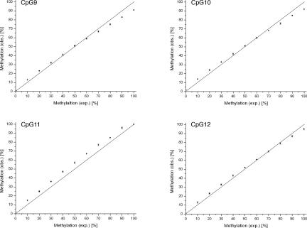
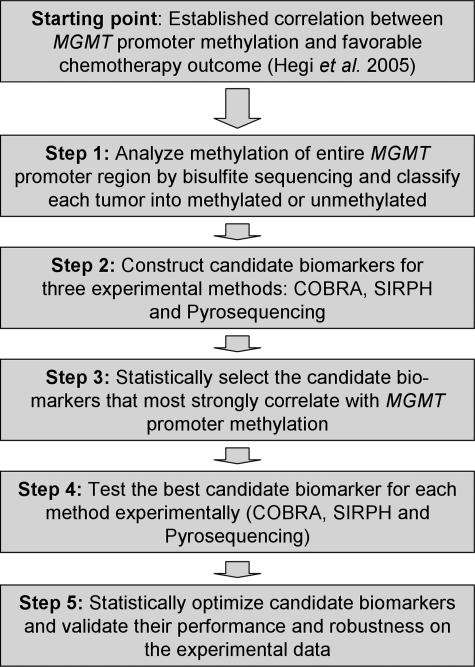
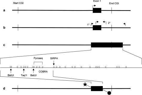
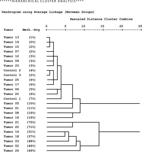
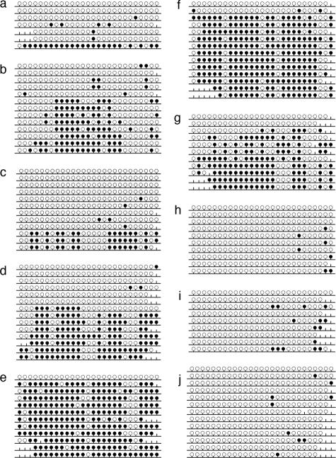
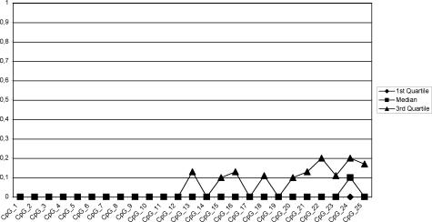
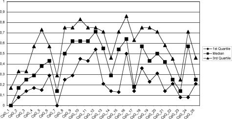
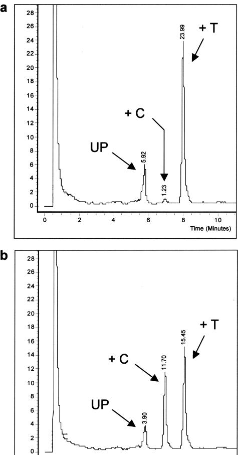
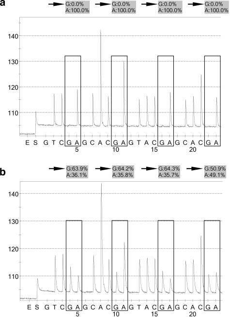
Resistance to chemotherapy is a major complication during treatment of cancer patients. Hypermethylation of the MGMT gene alters DNA repair and is associated with longer survival of glioblastoma patients treated with alkylating agents. Therefore, MGMT promoter methylation plays an important role as a predictive biomarker for chemotherapy resistance. To adopt this established correlation into a molecular diagnosis procedure, we compared and optimized three experimental techniques [combined bisulfite restriction analysis, a primer extension- and denaturing high-performance liquid chromatography-based method named SIRPH (SNuPE ion pair-reverse phase high-performance liquid chromatography), and pyrosequencing] with regard to their accuracy of detecting MGMT promoter methylation. Initially, bisulfite sequencing was used to obtain a comprehensive methylation profile of the MGMT promoter region in 22 glioblastoma samples and in three normal brain controls. Next, we statistically identified CpG sites that best discriminate between methylated and unmethylated MGMT promoters. These results were then used to design optimal combined bisulfite restriction analysis, SIRPH, and pyrosequencing assays for accurate and cost-efficient assessment of MGMT promoter methylation. We compared all three techniques with regard to their reliability and reproducibility on well-characterized tumor samples. The optimized pyrosequencing assay performed best and provides a sensitive, robust, and easy-to-use method for quantitative assessment of MGMT methylation, for both snap-frozen and paraffin-embedded specimens.


Figures

Figure 9
Figure 9
Titration curves obtained for each individual CpG position interrogated by the pyrosequencing assay. The SD of each data point is shown by a vertical error bar. The straight line reflects the theoretical curve. The titration experiment was performed as described in Materials and Methods. exp., expected; obs., observed.
Figure 1
Figure 1
Strategy for the optimization of MGMT promoter methylation detection as an epigenetic biomarker for chemotherapy resistance.
Figure 2
Figure 2
a: General map of the CpG island (CGI) spanning the promoter region and the first exon of the MGMT gene. b: Primer positions for the nested PCR approach used by Hegi et al. The second step (2°) used a methylation-sensitive primer established by Esteller et al. c: CpG map of the PCR product used for bisulfite sequencing of single clones, SIRPH, and pyrosequencing (biotin label not shown). d: Location of the double-labeled PCR product used for the COBRA assay.
Figure 3
Figure 3
Hierarchical clustering of samples by their MGMT promoter methylation profiles (methylation averages and standard deviations over all individual clones at CpG positions 1 to 25).
Figure 6
Figure 6
Methylation pattern obtained by bisulfite sequencing of single clones. a: Sample 08; b: sample 14; c: sample 16; d: sample 18; e: sample 21; f: sample 22; g: sample 24; h: sample 13; i: sample 20; and j: control sample 3. Filled circles correspond to methylated CpG positions, open circles correspond to unmethylated CpG positions, and the vertical lines without a circle correspond to CpG positions not determined in the sequence. The diagrams were generated with BiQ Analyzer software.
Figure 4
Figure 4
Methylation profile (methylation averages over all individual clones) of the low-methylation tumor subclass.
Figure 5
Figure 5
Methylation profile (methylation averages over all individual clones) of the high-methylation tumor subclass.
Figure 7
Figure 7
Typical chromatograms obtained in the SIRPH assay for an unmethylated sample (a: sample 15) and for a methylated sample (b: sample 21). UP, signal of the unextended primer; +C and +T, signal of the ddCTP and ddTTP extended primer, respectively. The signal seen in the beginning of the chromatogram is attributable to loading the sample on the column.
Figure 8
Figure 8
Typical pyrograms obtained for an unmethylated sample (a: sample 25) and for a methylated sample (b: sample 14). Each box represents one of four CpG positions interrogated by the pyrosequencing assay, starting on the left side with CpG 12 because of reverse sequencing the upper strand of the PCR product. As a consequence, a C/TpG position appears as a CpG/A. The incorporation of the base guanine in this context represents the methylated fraction (arrows), and incorporation of adenine represents the unmethylated fraction.
Figure 10
Figure 10
Marker scores for all three methods plotted against overall methylation level. The CO7 marker (a) is based on the analysis of CpGs 1, 2, 5, 8, and 9. The SI01 marker (b) is based on the analysis of CpG 13, and the Py15 marker (c) is based on the analysis of the CpG positions 9, 10, 11, and 12.

Similar articles

Cited by

References

    1. Ludlum DB. DNA alkylation by the haloethylnitrosoureas: nature of modifications produced and their enzymatic repair or removal. Mutat Res. 1990;233:117–126. - PubMed
    1. Pegg AE. Repair of O6-alkylguanine by alkyltransferases. Mutat Res. 2000;462:83–100. - PubMed
    1. Pegg AE, Dolan ME, Moschel RC. Structure, function, and inhibition of O6-alkylguanine-DNA alkyltransferase. Prog Nucleic Acid Res Mol Biol. 1995;51:167–223. - PubMed
    1. Wibley JEA, Pegg AE, Moody PCE. Crystal structure of the human O6-alkylguanine-DNA alkyltransferase. Nucleic Acids Res. 2000;28:393–401. - PMC - PubMed
    1. Karran P, Bignami M. DNA damage tolerance, mismatch repair and genome instability. Bioessays. 1994;16:833–839. - PubMed

Publication types

MeSH terms