. 2007 Jul;171(1):227-40.
doi: 10.2353/ajpath.2007.070182.

TDP-43 in familial and sporadic frontotemporal lobar degeneration with ubiquitin inclusions

Affiliations

TDP-43 in familial and sporadic frontotemporal lobar degeneration with ubiquitin inclusions

Nigel J Cairns et al. Am J Pathol. 2007 Jul.

Abstract

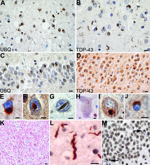
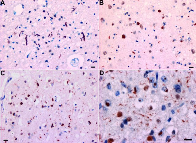
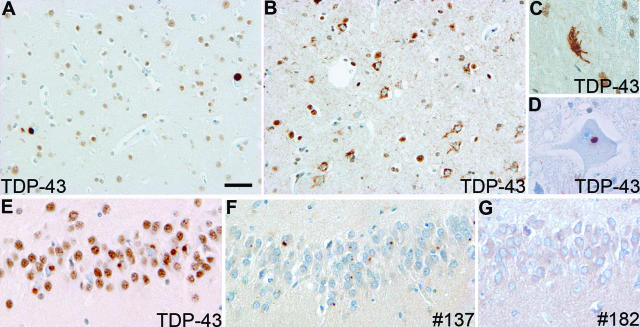
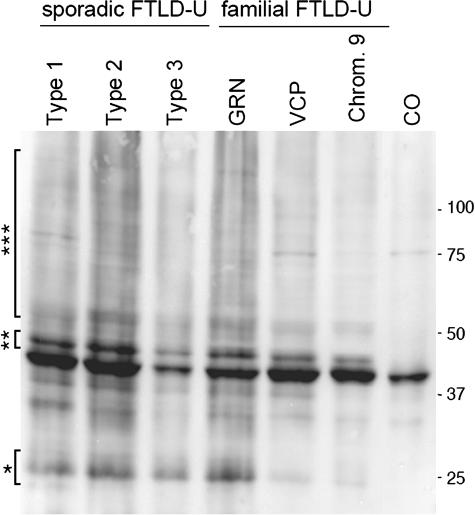
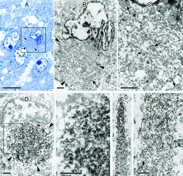
TAR DNA-binding protein 43 (TDP-43) is a major pathological protein of sporadic and familial frontotemporal lobar degeneration with ubiquitin-positive, tau-negative inclusions (FTLD-U) with or without motor neuron disease (MND). Thus, TDP-43 defines a novel class of neurodegenerative diseases called TDP-43 proteinopathies. We performed ubiquitin and TDP-43 immunohistochemistry on 193 cases of familial and sporadic FTLD with or without MND. On selected cases, immunoelectron microscopy and biochemistry were performed. Clinically defined frontotemporal dementias (FTDs) included four groups: 1) familial FTD with mutations in progranulin (n = 36), valosin-containing protein (n = 5), charged multivesicular body protein 2B (n = 4), and linked to chromosome 9p (n = 7); 2) familial cases of FTD with unknown gene association (n = 29); 3) sporadic FTD (n = 72); and 4) familial and sporadic FTD with MND (n = 40). Our studies confirm that the spectrum of TDP-43 proteinopathies includes most cases of sporadic and familial FTLD-U with and without MND and expand this disease spectrum to include reported families with FTD linked to chromosome 9p but not FTD with charged multivesicular body protein 2B mutations. Thus, despite significant clinical, genetic, and neuropathological heterogeneity of FTLD-U, TDP-43 is a common pathological substrate underlying a large subset of these disorders, thereby implicating TDP-43 in novel and unifying mechanisms of FTLD pathogenesis.


Figures

Figure 1
Figure 1
Spectrum of TDP-43 pathology in FTLD-U. Adjacent sections of superficial frontal neocortex showing NCIs, DNs, and isolated NIIs, stained for both ubiquitin (A) and TDP-43 (B). NCIs in the dentate granule cells stained for ubiquitin (C) and TDP-43 (D). Neuronal and glial inclusions include NCIs (E), round and lentiform NIIs (F and G); skein-like (H) and compact round (I) NCIs in lower motor neurons; and a glial cytoplasmic inclusion (J). Low-power micrograph showing numerous DNs in the hippocampus CA1 subfield (K). High-power micrograph showing a tortuous DN in a case of FTLD-U, subtype 1 (L). NCIs in the dentate fascia of a case of hippocampal sclerosis (M). A and C: Ubiquitin immunohistochemistry. B, D, EM: TDP-43 immunohistochemistry. Bars = 10 μm (AD and KM); 5 μm (EJ).
Figure 2
Figure 2
FTLD-U subtypes 1 to 4. A: Type 1 is characterized by long and tortuous DNs in laminae II/III with relatively few NCIs and no NII. B: Type 2 has numerous NCIs, relatively few DNs, and no NII. C: Type 3 has numerous NCIs and DNs and an occasional NII in lamina II. D: Type-4 pathology is characterized by numerous NIIs and DNs but few NCIs. TDP-43 immunohistochemistry. Bar = 10 μm.
Figure 3
Figure 3
Neuropathology of FTLD-U linked to chromosome 9p. TDP-43 immunohistochemistry showing compact TDP-43-positive cytoplasmic inclusions in neurons in the lower cortical layers (A); numerous neurons with diffuse granular cytoplasmic TDP-43 immunoreactivity in superficial frontal cortex (B); skein-like (C) and round (D) inclusion in motor neurons; and cytoplasmic inclusions in dentate granule cells (E). Note the absence of physiological, nuclear TDP-43 immunoreactivity in neurons with NCI. Inclusions in the dentate gyrus stained with monoclonal antibody 137 (F) but were negative with MAB 182 (G). Bars = 50 μm (A and B); 25 μm (CG).
Figure 4
Figure 4
Biochemical analysis of TDP-43. Immunoblot analysis probed with anti-TDP-43 antibody demonstrated a distinct pathological profile of TDP-43 with variable presence of pathological ∼25-kd bands (*), 45-kd bands (**), and high molecular smear (***) in all sporadic and familial FTLD-U brains (FTLD-U subtypes 1 to 3), including those with progranulin (GRN) and VCP mutations and those linked to chromosome 9p (Chrom. 9). Pathological TDP-43 bands were not detected in control brains (CO).
Figure 5
Figure 5
Fine structure of inclusions of FTLD-U. A: Light microscopy semithin section from the dentate gyrus of the hippocampus. Toluidine blue stain. Inset: Neuron with a round body (arrowheads) is adjacent to the nucleus. The nucleus is corrugated and indented compared with the other three normal neuronal nuclei. B and C: Re-embedded ultrathin section of the boxed cell in A and counterstained with uranyl acetate and bismuth subnitrate. B: Low-magnification electron micrograph showing a round body (arrowheads) of an NCI surrounded by mitochondria. C: Higher magnification of B shows an accumulation of granular and membranous material with a few mitochondria (arrows, mitochondria). D: Ultrathin section of a neuron from the dentate gyrus of the hippocampus, immunostained for TDP-43. An NCI (outlined by arrowheads) indenting the nucleus is labeled (no counterstain). E: Higher magnification of boxed area of D. F: Dystrophic neurite from the temporal lobe immunostained for TDP-43 (no counterstain). G: Same neurite as in F in an adjacent section counterstained with uranyl acetate and bismuth subnitrate. More filamentous material is seen than in the cell body of C, but no dense accumulations of filaments are present. Bars = 10 μm (A); 1 μm (BG).

References

    1. Neary D, Snowden JS, Mann DM. Classification and description of frontotemporal dementias. Ann NY Acad Sci. 2000;920:46–51. - PubMed
    1. Neary D, Snowden JS, Mann DM, Northen B, Goulding PJ, Macdermott N. Frontal lobe dementia and motor neuron disease. J Neurol Neurosurg Psychiatry. 1990;53:23–32. - PMC - PubMed
    1. Lipton AM, White CL, III, Bigio EH. Frontotemporal lobar degeneration with motor neuron disease-type inclusions predominates in 76 cases of frontotemporal degeneration. Acta Neuropathol (Berl) 2004;108:379–385. - PubMed
    1. Buratti E, Dork T, Zuccato E, Pagani F, Romano M, Baralle FE. Nuclear factor TDP-43 and SR proteins promote in vitro and in vivo CFTR exon 9 skipping. EMBO J. 2001;20:1774–1784. - PMC - PubMed
    1. Buratti E, Brindisi A, Pagani F, Baralle FE. Nuclear factor TDP-43 binds to the polymorphic TG repeats in CFTR intron 8 and causes skipping of exon 9: a functional link with disease penetrance. Am J Hum Genet. 2004;74:1322–1325. - PMC - PubMed

Publication types