Comparative Study
. 2007 Jul;171(1):338-48.
doi: 10.2353/ajpath.2007.060589.

Radiogenic lymphangiogenesis in the skin

Affiliations
Comparative Study

Radiogenic lymphangiogenesis in the skin

Susanne Jackowski et al. Am J Pathol. 2007 Jul.

Abstract

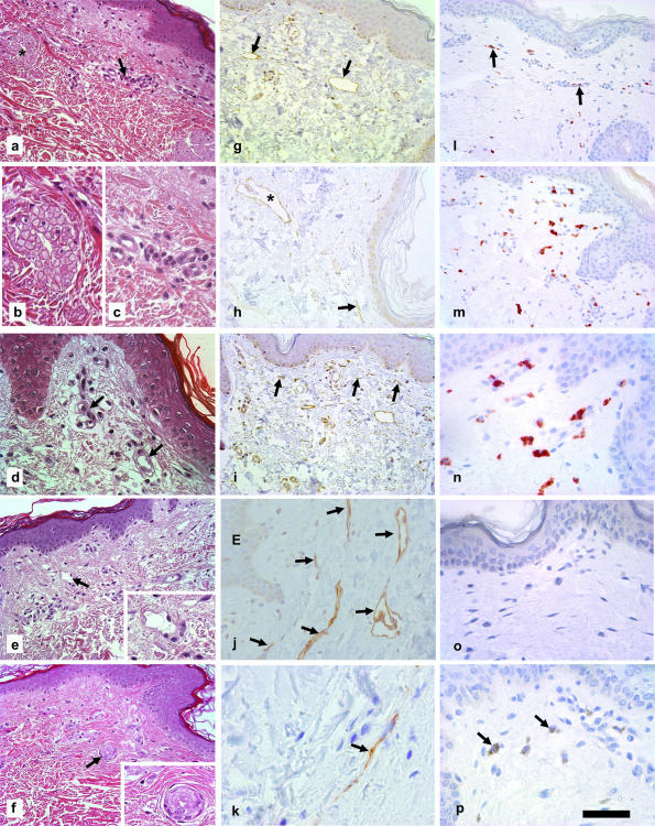
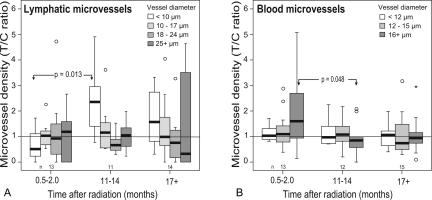
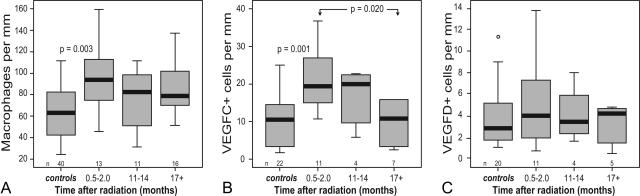
The time course of microvascular changes in the environment of irradiated tumors was studied in a standardized human protocol. Eighty skin biopsies from 40 patients with previously treated primary breast cancer were taken from irradiated skin and corresponding contralateral unirradiated control areas 2 to 8 weeks, 11 to 14 months, or 17+ months after radiotherapy (skin equivalent dose 30 to 40 Gy). Twenty-two biopsies of 11 melanoma patients who had undergone lymph node dissection were used for unirradiated control. We found an increase of total podoplanin(+) lymphatic microvessel density resulting mainly from a duplication of the density of smallest lymphatic vessels (diameter <10 microm) in the samples taken 1 year after radiation. Our findings implicate radiogenic lymphangiogenesis during the 1st year after therapy. The numbers of CD68(+) and vascular endothelial growth factor-C(+) cells were highly elevated in irradiated skin in the samples taken 2 to 8 weeks after radiotherapy. Thus, our results indicate that vascular endothelial growth factor-C expression by invading macrophages could be a pathogenetic route of induction of radiogenic lymphangiogenesis.


Figures

Figure 1
Figure 1
Microphotographs of the upper dermal plexus of irradiated and naive control skin. a–f: H&E staining. a: Four weeks after radiation, sparse perivascular mononuclear infiltrate (arrow) and shrunken sebaceous glands (asterisk) were evident. b and c: Details of a (compare with asterisk or arrow). d: Greater enlargement shows microvessel degeneration (arrows). e: Twelve months after radiation, lymphatic spaces (arrow, inset), and disfigured microvessels were visible. f: Similar microvessel changes and, in this case, sweat duct changes (inset) could be found 23 months after radiotherapy. g–k: Podoplanin immunostaining (hematoxylin counterstained). g: In periaxillar normal control skin, small and large lymphatic vessels (arrows) can be found regularly in horizontal orientation. h: Four weeks after radiation, lymphatic vessels (asterisk) showed vertical enlargement of the lumina, but the number of small lymphatics (arrow) was comparable with the controls. i: One year after radiotherapy, the number of small horizontally oriented lymphatics (>10 μm diameter, arrows) was increased dramatically. j and k: Higher enlargement of podoplanin staining 11 months (j) or 17 months (k) after radiation: Only clearly identifiable podoplanin+ longitudinal structures were evaluated as lymphatics (arrows; E, epidermis). l–n: CD68 immunostaining (red). l: Low density of CD68+ cells (macrophages, arrows) in the normal control. m and n: High densities of CD68+ cells were characteristic 4 weeks after radiation. o and p: VEGF-C immunostaining (brown). Normal control (o) and irradiated skin (4 weeks after radiation) (p) showed significant difference in the densities of VEGF-C+ cells (arrows). Scale bars: 100 μm (a, e–i, l, and m); 50 μm (b–d, j, n–p, and insets in e and f); 25 μm (k).
Figure 2
Figure 2
Ratios of “treated/control” densities of different microvessel calibers during different time stages of the postradiation period. a: Lymphatics with four groups of microvessel diameter and three time groups after radiotherapy, respectively. b: Analogous figure of blood microvessel densities with three groups of microvessel diameter ranges. Intertime group significance analysis by U-test. Boxes include all values from 25th to 75th percentile; –, median; 𝕋⊥, largest to smallest observed values that are not outliers; ○, outlier more than 1.5 box lengths from the 25 or 75 percentile; and asterisk, outlier value >2 × SD from mean.
Figure 3
Figure 3
Densities of CD68+ cells (a), VEGF-C+ (b), or VEGF-D+ (c) cells. Dependence on the time after radiation and comparison with the data of unirradiated control skin. Wilcoxon test, comparison of controls with irradiated samples; U-test, between irradiated samples. Boxes include all values from 25th to 75th percentile; –, median; 𝕋⊥, largest to smallest observed values that are not outliers; ○, outlier more than 1.5 box lengths from the 25 or 75 percentile.

Similar articles

Cited by

References

    1. Archambeau JO, Ines A, Fajardo LF. Correlation of the dermal microvasculature morphology with the epidermal and the endothelial population changes produced by single X ray fractions of 1649, 2231 and 2619 rad in swine. Int J Radiat Oncol Biol Phys. 1985;11:1639–1646. - PubMed
    1. Takahashi S, Sugimoto M, Kotoura Y, Sasai K, Oka M, Yamamuro T. Long-term changes in the haversian systems following high-dose irradiation: an ultrastructural and quantitative histomorphological study. J Bone Joint Surg Am. 1994;76:722–738. - PubMed
    1. Tsai JH, Makonnen S, Feldman M, Sehgal CM, Maity A, Lee WM. Ionizing radiation inhibits tumor neovascularization by inducing ineffective angiogenesis. Cancer Biol Ther. 2005;4:1395–1400. - PubMed
    1. Fajardo LF. The pathology of ionizing radiation as defined by morphologic patterns. Acta Oncol. 2005;44:13–22. - PubMed
    1. Reinhold HS. The influence of radiation on blood vessels and circulation. Chapter IV. Structural changes in blood vessels. Curr Top Radiat Res Q. 1974;10:58–74. - PubMed

Publication types

MeSH terms

Substances