. 2007 Jul;171(1):349-60.
doi: 10.2353/ajpath.2007.060831.

Myeloperoxidase interacts with endothelial cell-surface cytokeratin 1 and modulates bradykinin production by the plasma Kallikrein-Kinin system

Affiliations

Myeloperoxidase interacts with endothelial cell-surface cytokeratin 1 and modulates bradykinin production by the plasma Kallikrein-Kinin system

Joshua M Astern et al. Am J Pathol. 2007 Jul.

Abstract

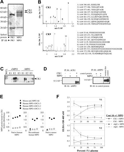
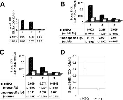
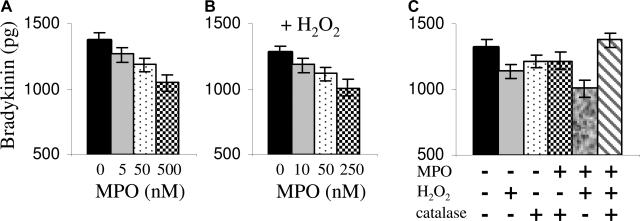
During an inflammatory state, functional myeloperoxidase (MPO) is released into the vessel as a result of intravascular neutrophil degradation. One mechanism of resulting cellular injury involves endothelial internalization of MPO, which causes oxidative damage and impairs endothelial signaling. We report the discovery of a protein that facilitates MPO internalization, cytokeratin 1 (CK1), identified using affinity chromatography and mass spectrometry. CK1 interacts with MPO in vitro, even in the presence of 100% human plasma, thus substantiating biological relevance. Immunofluorescent microscopy confirmed that MPO added to endothelial cells can co-localize with endogenously expressed CK1. CK1 acts as a scaffolding protein for the assembly of the vasoregulatory plasma kallikrein-kinin system; thus we explored whether MPO and high molecular weight kininogen (HK) reside on CK1 together or whether they compete for binding. The data support cooperative binding of MPO and HK on cells such that MPO masked the plasma kallikrein cleavage site on HK, and MPO-generated oxidants caused inactivation of both HK and kallikrein. Collectively, interactions between MPO and the components of the plasma kallikrein-kinin system resulted in decreased bradykinin production. This study identifies CK1 as a facilitator of MPO-mediated vascular responses and thus provides a new paradigm by which MPO affects vasoregulatory systems.


Figures

Figure 1
Figure 1
CK1 and CK9 specifically bind MPO. A: Coomassie-stained gel of immunopurified MPO or proteinase 3 (PR3) complexes resolved by SDS-PAGE. Mass spectrometry analysis of the unique bands in the MPO-spiked membrane fraction identified CK1 and CK9. The spectra are shown in B. The tryptic peptides are numbered and displayed with the corresponding sequences. C: Western blot analysis verified that CK1 binds to MPO. HUVEC whole cell lysates were incubated with purified MPO and passed over a monoclonal MPO-specific antibody immobilized on Aminolink Plus coupling gel. The proteins eluted from the beads were subjected to SDS-PAGE and Western blot analysis using CK1-specific antibody. E1, E2, and E3 are sequential elution fractions from the beads. D: In vitro mixing of purified proteins resulted in the binding of MPO to CK1 but not to a control recombinant protein (BMP7). Western blot analysis (IB) of immunopurified complexes (IP) indicated that an anti-CK1 antibody (Ab) immunoprecipitated MPO but only when complexed with CK1. MPO does not bind the control recombinant protein demonstrating the specificity of the MPO-CK1 interaction. E: Sandwich ELISAs: the different shapes represent the antibody used to detect MPO binding. Solid black indicates the ELISA well was coated with a CK1 antibody, and open shape indicates the well was coated with normal IgG. Antibody-captured CK1 was incubated with purified MPO, recombinant MPO, or purified mouse MPO. Results show that purified MPO was detected by the monoclonal MPO antibody, but this antibody did not react with recombinant MPO or mouse MPO. Anti-MPO antibodies from three patients with anti-neutrophil cytoplasmic autoantibody disease (MPO-ANCA) recognized the CK1-MPO complex with all three forms of MPO protein. F: CK1 captures MPO directly from human plasma. ELISA wells were coated with either anti-CK1 antibody or normal IgG. CK1 was then captured and incubated with or without MPO in increasing amounts of plasma drawn from two healthy volunteers (V1 and V2).
Figure 2
Figure 2
MPO and CK1 co-localize in living cells. A: EA.hy926 cells were incubated with MPO in serum-free medium. MPO was detected with the DAKO mouse anti-MPO antibody, and CK1 was detected using a polyclonal anti-CK1 antibody. The yellow foci in the merged image indicate areas of co-localized CK1 and MPO. B and C: HUVECs were also exposed to MPO in serum-free (B) or serum-containing (C) medium. MPO was labeled in this experiment with a mouse MPO antibody from Abcam. CK1 was labeled with the same polyclonal antibody used in A. Widespread co-localization was noted in both conditions. D: Negative control for the CK1 label on HUVECs (anti-rabbit secondary antibody only). E: Negative control for the MPO label on HUVECs (cells not treated with MPO but exposed to the anti-MPO antibody and its corresponding secondary antibody). Original magnifications, ×60.
Figure 3
Figure 3
Internalization of MPO by endothelial cells is reduced by blocking the MPO and CK1 interaction. Experiments are flow cytometry analyses of endothelial cells labeled with a polyclonal anti-MPO antibody. A: EA.hy926 cells were pretreated with a CK1-blocking mix consisting of factor XII (FXII) and a goat anti-CK1 antibody (CK Ab) raised against a kininogen-binding site of CK1. Cells were then exposed to 2 or 5 μg/ml MPO for 10 minutes. B: HUVECs were pretreated with a pan-CK (pCK Ab) antibody and then 2 μg/ml MPO was added for 10 minutes. C: EA.hy926 cells were pretreated with either a pan-CK (pCK Ab) antibody or normal mouse IgG before 0.5 μg/ml MPO was added for 10 minutes. FITC, fluorescein isothiocyanate; FSC, forward scatter.
Figure 4
Figure 4
MPO enhances kininogen binding to endothelial cells and the proteins co-localize on the endothelial cell surface and intracellularly. A: MPO enhances kininogen binding. HUVECs were treated with biotinylated high molecular weight kininogen (bHK) either alone or in the presence of a 50-fold molar excess of unlabeled high molecular weight kininogen (uHK) or MPO for 30 minutes at 37°C. Bound bHK was detected using alkaline phosphatase-conjugated streptavidin. Unlabeled HK competed with bHK binding resulting in 50% less bHK protein bound. MPO enhanced bHK binding by 5.35-fold. BE: MPO and kininogen co-localize in/on endothelial cells. HUVECs were either treated with bHK and purified MPO (B–D) or protein-free medium. The cells were labeled using both Alexa Fluor 488-conjugated streptavidin (B) and an anti-MPO antibody with corresponding Alexa Fluor 568 secondary antibody (C). D: The merged image of B and C showing a high degree of co-localization between MPO and bHK. The merged image of cells that were treated with protein-free medium is shown in E. Original magnifications, ×60.
Figure 5
Figure 5
MPO binds kininogen. A: Direct ELISA: column 1: biotinylated kininogen (bHK) binds immobilized MPO but not bovine serum albumin as detected by alkaline phosphatase-conjugated streptavidin; column 2: bHK binding is reduced in the presence of excess unlabeled kininogen (uHK); column 3: unlabeled kininogen alone was negative. B: Sandwich ELISA: further validation of the direct interaction between MPO and kininogen. Column 1: monoclonal MPO antibody-captured MPO binds bHK as detected by alkaline phosphatase-conjugated streptavidin; column 2: competition of bHK binding in the presence of excess uHK; column 3: unlabeled kininogen alone was negative. Shown are the averaged results of three independent trials ± SDs. C: Sandwich ELISA: validation using a second MPO antibody. Column 1: polyclonal MPO antibody-captured MPO binds bHK as detected by alkaline phosphatase-conjugated streptavidin; column 2: competition of bHK binding in the presence of excess uHK; column 3: unlabeled kininogen alone was negative. Shown are the results of three independent trials ± SDs. D: MPO captures bHK from plasma. HK-depleted plasma was replenished with bHK to physiological levels. Immobilized MPO was used as bait to capture bHK from plasma. Shown are the results (with SE bars) of three independent experiments.
Figure 6
Figure 6
MPO interferes with the plasma kallikrein-kininogen system: bradykinin production is diminished. A: MPO interfered with bradykinin production through steric hindrance at high concentrations. Shown are the results of four independent experiments measuring the bradykinin liberated from 50 nmol/L HK in the presence of MPO. B: Provided with its substrate (H2O2), MPO affects bradykinin production at lower concentrations. Shown are the results of three independent experiments. C: The enzymatic activity of MPO (conversion of H2O2 to HOCl) hinders bradykinin production, which is rescued in the presence of catalase. Shown are the results of three independent experiments.
Figure 7
Figure 7
Hypochlorous acid (HOCl) inhibits bradykinin production. A: The proteolytic activity of kallikrein is abolished by HOCl dose-dependently but is protected when HOCl is quenched with l-methionine (20-fold molar excess). Shown are the results (with SDs) of three separate experiments. B: HK is oxidized by HOCl, resulting in a product uncleavable by active kallikrein. HK was pretreated with increasing concentrations of HOCl. A 20-fold excess of l-methionine was added (20 minutes) before the addition of active kallikrein. Bradykinin production was assessed by ELISA. Shown are the results from four separate experiments. C: HOCl inactivates kininogen by oxidizing a critical methionine residue. HOCl-oxidized HK was treated with a methionine sulfoxide reductase (PilB), and the inhibition of bradykinin production was reversed. DTT, dithiothreitol.
Figure 8
Figure 8
Proposed schematic for the interactions of MPO with endothelial cells and the plasma kallikrein-kininogen system. When a neutrophil releases its granule constituents and oxygen radicals at sites of inflammation, MPO can leak into the lumen of the vessel. A: Endothelial cells bind and internalize MPO, in part through interactions with the cell-surface protein CK1; MPO and CK1 enter the cells in complex. B: MPO can also enter cells through other mechanisms that have yet to be fully characterized. C–F: MPO can modulate the action of the plasma kallikrein-kininogen system. c: MPO associates directly with high molecular weight kininogen (HK) and increases the amount of HK bound to the cells; this complex appears to internalize. D: When MPO and kininogen are coupled, kallikrein is unable to cleave HK. MPO uses the hydrogen peroxide generated during a neutrophil’s respiratory burst to oxidize chloride and produce hypochlorous acid (represented by the yellow asterisk). E: Hypochlorous acid can oxidize and inactivate HK by altering kallikrein’s cleavage site (F), as well as abrogate the protease activity of kallikrein. G: In the absence of MPO, endothelial surface proteins including CK1 bind circulating high molecular weight kininogen. Plasma kallikrein subsequently cleaves bradykinin (small red circle with B) from kininogen; bradykinin then binds to specific endothelial receptors to induce nitric oxide generation.

References

    1. Nauseef WM, Malech HL. Analysis of the peptide subunits of human neutrophil myeloperoxidase. Blood. 1986;67:1504–1507. - PubMed
    1. Pullar JM, Vissers MC, Winterbourn CC. Living with a killer: the effects of hypochlorous acid on mammalian cells. IUBMB Life. 2000;50:259–266. - PubMed
    1. Arimura Y, Minoshima S, Kamiya Y, Tanaka U, Nakabayashi K, Kitamoto K, Nagasawa T, Sasaki T, Suzuki K. Serum myeloperoxidase and serum cytokines in anti-myeloperoxidase antibody-associated glomerulonephritis. Clin Nephrol. 1993;40:256–264. - PubMed
    1. Baldus S, Heeschen C, Meinertz T, Zeiher AM, Eiserich JP, Munzel T, Simoons ML, Hamm CW. Myeloperoxidase serum levels predict risk in patients with acute coronary syndromes. Circulation. 2003;108:1440–1445. - PubMed
    1. Brennan ML, Penn MS, Van Lente F, Nambi V, Shishehbor MH, Aviles RJ, Goormastic M, Pepoy ML, McErlean ES, Topol EJ, Nissen SE, Hazen SL. Prognostic value of myeloperoxidase in patients with chest pain. N Engl J Med. 2003;349:1595–1604. - PubMed

Publication types

MeSH terms