. 2007 Jul;130(1):21-40.
doi: 10.1085/jgp.200609729.

Physiological properties of rod photoreceptor cells in green-sensitive cone pigment knock-in mice

Affiliations

Physiological properties of rod photoreceptor cells in green-sensitive cone pigment knock-in mice

Keisuke Sakurai et al. J Gen Physiol. 2007 Jul.

Abstract

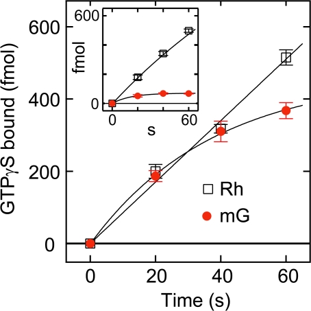
Rod and cone photoreceptor cells that are responsible for scotopic and photopic vision, respectively, exhibit photoresponses different from each other and contain similar phototransduction proteins with distinctive molecular properties. To investigate the contribution of the different molecular properties of visual pigments to the responses of the photoreceptor cells, we have generated knock-in mice in which rod visual pigment (rhodopsin) was replaced with mouse green-sensitive cone visual pigment (mouse green). The mouse green was successfully transported to the rod outer segments, though the expression of mouse green in homozygous retina was approximately 11% of rhodopsin in wild-type retina. Single-cell recordings of wild-type and homozygous rods suggested that the flash sensitivity and the single-photon responses from mouse green were three to fourfold lower than those from rhodopsin after correction for the differences in cell volume and levels of several signal transduction proteins. Subsequent measurements using heterozygous rods expressing both mouse green and rhodopsin E122Q mutant, where these pigments in the same rod cells can be selectively irradiated due to their distinctive absorption maxima, clearly showed that the photoresponse of mouse green was threefold lower than that of rhodopsin. Noise analysis indicated that the rate of thermal activations of mouse green was 1.7 x 10(-7) s(-1), about 860-fold higher than that of rhodopsin. The increase in thermal activation of mouse green relative to that of rhodopsin results in only 4% reduction of rod photosensitivity for bright lights, but would instead be expected to severely affect the visual threshold under dim-light conditions. Therefore, the abilities of rhodopsin to generate a large single photon response and to retain high thermal stability in darkness are factors that have been necessary for the evolution of scotopic vision.


Figures

Figure 1.
Figure 1.
Generation of mouse green cone pigment knock-in mice. (A) Targeting strategy to generate mouse green knock-in mice. The restriction maps of the mouse rhodopsin genomic locus (top), the targeting vector containing mouse green opsin cDNA (mG) (middle) and the predicted genomic structure of the knock-in allele after homologous recombination (bottom). The five exons of the rhodopsin gene are shown as the open rectangles (1–5). The crossed broken lines indicate the areas of homology between the wild-type rhodopsin locus and the targeting vector. The chimeric intron is inserted into XhoI site in the 5′ untranslated region of rhodopsin gene. The translation initiation codon of mouse green opsin cDNA sequence (mG) was placed at the same position as that of mouse rhodopsin gene. The subsequent PA represents the rhodopsin polyadenylation signals. The phosphoglycerate kinase driven neomycin resistance gene (neo) is flanked by 34-bp loxP sequences (triangles). Digestions with BamHI distinguish the homologous recombinant fragments (7 kb) from the wild-type fragments (11 kb). Restriction sites: B, BamHI; C, ClaI; E, EcoRI; S, SpeI; X, XhoI. (B) Southern blots of BamHI-digested mouse tail genomic DNA from wild-type (Rh/Rh), heterozygous (Rh/mG), and homozygous (mG/mG) mice. The 11-kb fragments from wild-type allele and 7-kb fragments from targeted allele are detected by the 5′ flanking probe shown in A. (C) RT-PCR analysis to verify the replacement of the transcripts. The upper diagram represents the predicted RT-PCR products from rhodopsin and introduced mouse green transcripts. The length of RT-PCR products derived from wild-type and knock-in alleles are 1306 and 1348 bp, respectively. SacI digestion yields 950- and 356-bp fragments of RT-PCR product from rhodopsin transcripts. On the other hand, SmaI digestion yields 810- and 538-bp fragments from introduced mouse green transcripts. In wild-type lanes (Rh/Rh), SacI digestion yielded two fragments of 950 and 356 bp, but SmaI did not. Conversely, in mG/mG lanes, SmaI digestion yielded two fragments of 810 and 510 bp, but SacI digestion did not. In the Rh/mG lanes, both Smal and Sacl digestion yielded two fragments reflecting the presence of both rhodopsin and mouse green transcripts. In the negative control (N.C.) lane, no PCR product was detected from the total RNA sample of mG/mG retina, ensuring that PCR products observed here were derived from mRNA. M represents DNA size marker (from top to bottom: 1,517, 1,200, 1,000, 900, 800, 700, 600, 500, 400, and 300 bp). (D) Western blots using the antibodies against rhodopsin (top panel) and mouse green (bottom panel). Rhodopsin was detected by rhodopsin 1D4 antibody at 38-kD position, which showed the genotype-dependent expression. A faint band was detected for mouse green endogenously expressed in cone photoreceptors at 42 kD in the Rh/Rh lane (arrow), whereas in the Rh/mG and mG/mG lanes intense bands from the recombinant mouse green were detected at the same position. An equal amount of retinal lysate (0.003% of a retina for rhodopsin 1D4 antibody and 1.3% of a retina for anti-mouse green antibody) was loaded in each lane.
Figure 2.
Figure 2.
Expression of the functional proteins in the knock-in mice. (A) Photosensitivity measurements of rhodopsin (open squares) and mouse green (red closed circles) at 500 nm. The amount of residual pigment was plotted on a logarithmic scale against the numbers of incident photons. By comparing the bleaching rate (photons−1 μm2) of the fitted exponential curves, which was (8.0 ± 0.3) × 10−18 for rhodopsin and (7.9 ± 0.3) × 10−18 for mouse green, the ratio of the photosensitivity at 500 nm of mouse green to that of rhodopsin was estimated to be 0.99. Errors indicate 95% confidence interval of the parameters. (B) Estimation of the ratio of the amount of rhodopsin and mouse green in Rh/mG retinas. The visual pigments extracted from the retinas of wild-type (open squares), Rh/mG (open triangles), and mG/mG (red closed circles) mice were subjected to the sensitivity measurements in 200 mM neutralized hydroxylamine at 20°C. The remaining visual pigments (%) were plotted as a function of the incubation time (h). The bleaching processes of rhodopsin (open squares) and mouse green (red closed circles) were fit with single-exponential functions with time constants of 127 h and 1.93 h, respectively. The bleaching process of a mixture of rhodopsin and mouse green (open triangle) in heterozygous retinas monitored at 500 nm was fit with a double- exponential function with time constants of 127 h (89%) and 1.93 h (11%). After correcting for the different molecular extinction coefficient at 500 nm, the proportion of rhodopsin to mouse green was estimated to be 90 to 10. (Inset) The difference absorption spectra of the Rh/mG retinal extract. Curve 1 is the difference spectrum calculated by subtracting the spectrum immediately after adding hydroxylamine from that 4 h after incubation in the presence of hydroxylamine. Curve 2 is the difference spectrum calculated by subtracting the spectrum 4 h after incubation in the presence of neutralized hydroxylamine from that after complete bleaching by irradiation with >500-nm light. (C) Comparison of the phototransduction proteins in retinal homogenates. (Left) Western blots of wild-type (Rh/Rh), heterozygous (Rh/mG), and homozygous (mG/mG) mice. In each lane, 1.3% homogenate of one retina at 4-wk-old mice were electrophoresed for blotting. (Right) Protein expression levels in the retinas of homozygous (mG/mG) mice relative to those of wild-type mice. Mice of 3, 4, and 6 wk old were subjected to the experiments. The horizontal broken line indicates the ratio of total surface area of outer segment of mG/mG rod relative to that of wild-type one. The values from three to four experiments were averaged with SEM bars. Gt1α, rod transducin α subunit; PDEα, phosphodiesterase α subunit; PDEβ, phosphodiesterase β subunit; CNG1, cyclic nucleotide-gated channel 1; GRK1, G-protein receptor kinase 1.
Figure 3.
Figure 3.
Morphology and histological expression patterns of rhodopsin and mouse green. Retinal sections of 3-wk-old wild-type, heterozygous, and homozygous mice are shown in A–D, E–H, and I–L. (A, E, and I) Nomarski images. (B, F, and J) Stained for rhodopsin (green). (C, G, and K) Stained for mouse green (red). (D, H, and L) Merged. OS, outer segments; ONL, outer nuclear layer; OPL, outer plexiform layer; INL, inner nuclear layer. (M–P) Electron micrographs of the photoreceptors of 3-wk-old (M) wild-type, (N) heterozygous, and (O) homozygous mice. Wild-type discs stacked tightly within the ROS and were oriented perpendicular to the long axis of the ROS. Heterozygous (Rh/mG) discs appeared to be normal. Homozygous (mG/mG) discs of normal size stacked neatly at the basal region of outer segments but the distal discs occasionally formed vesicle-like structures (arrowheads). (P) Photoreceptors of 7-wk-old homozygous (mG/mG) mouse. Magnification bars are 20 μm (A–L) and 1 μm (M–P).
Figure 4.
Figure 4.
Comparison of G-protein activation by rhodopsin (open squares) and mouse green (red filled circles) in ROS membranes at 0°C. The amount of light-dependent GTPγS-bound transducin was plotted as a function of incubation time after irradiation. The solid curves indicate the results by fitting a linear function for the data of rhodopsin and a single-exponential function for the data of mouse green. The initial rate of GTPγS bound (fmol s−1) was 10 ± 0.4 for rhodopsin (n = 4) and 13 ± 1.3 for mouse green (n = 4; P > 0.05). (Inset) Time courses of G-protein activation efficiencies of rhodopsin (open squares) and mouse green (red filled circles) in ROS membrane at 37°C. The initial rate of GTPγS bound (fmol s−1) was 8.3 ± 0.3 for rhodopsin (n = 4) and 4.6 ± 0.7 for mouse green (n = 3; P < 0.05). Error bars indicate SEM.
Figure 5.
Figure 5.
Single-cell recordings of wild-type (Rh/Rh) and homozygous (mG/mG) rods. (A) Normalized flash responses of the wild-type (top) and the mG/mG (bottom) rods to increasing 500-nm flash strength delivered at time 0 with 20 ms duration. Each trace is the averaged responses from multiple flash trials. Flash monitor is shown by bottom trace in each panel. Flash intensities were 2.5, 4.9, 10, 19, 43, 82, 170, 330, and 730 photons μm−2 in the wild-type rod, while 43, 82, 170, 330, 730, 1.4 × 10³, 2.9 × 10³, 5.6 × 10³, and 1.2 × 10⁴ photons μm−2 in the mG/mG rod. The maximal responses were 9.4 pA for the wild-type rod and 8.4 pA for the mG/mG rod. (B) Normalized peak responses plotted as a function of photoisomerizations per rod. Responses of wild-type (open squares; n = 22) and mG/mG (red filled circles; n = 14) rods were fitted with: r/rmax = 1 − exp (−k'Φ), where Φ is photoisomerizations defined as Eq. 3. In(2)/k' provides photoisomerizations giving the half-saturating response. Mean half-saturating photoisomerizations collected from 22 wild-type and 14 mG/mG rods were 9.7 R* and 39 R*, respectively. (C) Amplification constants as a function of photoisomerizations per flash. Each point of wild-type (open squares; n = 13) or mG/mG (red filled circles; n = 6) rods is the mean value of A, which was determined by fitting with Eq. 2 to the rising phase of the normalized response. Amplification constant, A0, of each cell was estimated by fitting with: A = A0/(1 + Φ/Φ0), where A0 is the constant value of A at lower intensities and Φ0 is the photoisomerizations at which A declines to the half maximum. The mean values of A0 were 25 s−2 from 13 wild-type rods and 7.0 s−2 from six mG/mG rods. Error bars indicate SEM.
Figure 6.
Figure 6.
Characterization of the E122Q rhodopsin and mouse green in the heterozygous (RhEQ/mG) rods. (A) Estimation of the ratio of the amount of E122Q rhodopsin and mouse green in RhEQ/mG retinas. The pigments extracted from the retinas of RhEQ/RhEQ (blue open squares), RhEQ/mG (closed triangle), and mG/mG (red closed circles) mice were subjected to the sensitivity measurements in 200 mM neutralized hydroxylamine at 20°C. The remaining visual pigments (%) were plotted as a function of the incubation time (h). The bleaching processes of E122Q rhodopsin and mouse green were fit with single-exponential functions with time constants of 107 h and 1.93 h, respectively. The bleaching process of a mixture of E122Q rhodopsin and mouse green from heterozygous mice was monitored at 498 nm (isosbestic point of E122Q rhodopsin and mouse green) and then fit with a double-exponential function with time constants of 107 h (91%) and 1.93 h (9%). The proportion of E122Q rhodopsin to mouse green was estimated to be 91 to 9. (Inset) The difference absorption spectra of the RhEQ/mG retinal extract. Curve 1 is the difference spectrum calculated by subtracting the spectrum immediately after adding hydroxylamine from that 4 h after incubation in the presence of hydroxylamine. Curve 2 is the difference spectrum calculated by subtracting the spectrum 4 h after incubation in the presence of neutralized hydroxylamine from that after complete bleaching by irradiation with >500-nm light. (B) Spectral sensitivities at 600, 652, and 676 nm relative to 500 nm. Mean spectral sensitivities at these wavelengths relative to that at 500 nm, which were obtained from RhEQ/RhEQ (blue open squares; n = 10), mG/mG (red closed circles; n = 11), and RhEQ/mG (closed triangles; n = 14) rods by suction electrode recordings, were plotted against wavelength. The values are described in Table II. The solid curves are the absorption spectra (at 20°C) of E122Q rhodopsin (blue curve; λmax = 487 nm) and mouse green (red curve; λmax = 510 nm), which are normalized at 500 nm. The broken curves are the template spectra of visual pigments whose λmax are located at 487 nm (blue broken curve) and 510 nm (red broken curve). These template spectra were described previously (Govardovskii et al., 2000). (Inset) Photosensitivity measurements of E122Q rhodopsin (blue open squares) and mouse green (red closed circles) at 500 nm. The amounts of residual pigment were plotted on a logarithmic scale against the numbers of incident photons. By comparing the bleaching rate (photons−1 μm2) of the fitted exponential curves, which was (7.3 ± 0.2) × 10−18 for E122Q rhodopsin and (7.9 ± 0.3) × 10−18 for mouse green, the ratio of photosensitivity at 500 nm of E122Q rhodopsin to that of mouse green was estimated to be 0.93. Errors indicate 95% confidence interval of the parameters. (C) The grand mean of dim flash response kinetics from 14 heterozygous (RhEQ/mG) rods evoked by consecutive identical flashes with 500 nm (black trace) and 676 nm (gray trace). These traces are normalized at peak response. (Inset) The variance-to-mean ratios of the responses evoked by 500-nm (black bar) and 676-nm (gray bar) flashes. These values are the averages from 14 RhEQ/mG rods. The difference in the ensemble variance-to-mean ratio is significant between 500-nm and 676-nm light (P < 0.005; two-tailed paired Student's t test). Error bars indicate SEM.
Figure 7.
Figure 7.
Noise analyses in wild-type and mG/mG rods. (A) Typical traces of membrane currents of wild-type rod in the dark (two top traces) and in the presence of saturating lights (two bottom traces) that close all channels in rod. It should be noted that, in wild-type rods, dark current after bleaching of visual pigments did not recover. The intensity of the saturating 500-nm light was 3.8 × 10⁴ photons μm−2 s−1. The leak resistance was 7.5 MΩ and circulating current was 8.8 pA. (B) Typical traces of membrane currents of mG/mG rod in the dark (two top traces), in the presence of saturating lights (two middle traces), and in darkness after bleaching >99% visual pigments by light irradiation (two bottom traces). The intensity of the saturating 500-nm light was 6.6 × 10⁵ photons μm−2 s−1. The leak resistance was 10.6 MΩ and circulating current was 9.7 pA. (C) Power spectra of current fluctuations calculated from the data of eight wild-type rods in the dark (closed circles) and those in the presence of saturating lights (open circles). The horizontal dotted line denotes the mean Johnson noise level according to the Nyquist equation. The value was 0.0021 pA2 Hz−1 (n = 8). (D) Power spectra calculated from the data of 16 mG/mG rods in the dark (closed circles), those in the presence of saturating light (open circle), and those in the dark after bleaching >99% visual pigment (open squares). The horizontal dotted line denotes the mean Johnson noise level according to the Nyquist equation. The value was 0.0021 pA2 Hz−1 (n = 16). (E) The recovery of circulating current after bleaching mouse green to various extents in mG/mG rods. The post-bleach circulating currents scaled to the prebleach current were plotted against the amount of remaining pigments. Open triangles indicate the individual data obtained from each cell and closed triangles indicate the data averaged from individual data for given remaining pigments. (F) Difference power spectra derived from mG/mG rods (n = 16). The mean difference spectrum calculated by subtracting the power spectrum obtained in the presence of saturating lights from that obtained in darkness is shown as closed circles. The solid curve was drawn according to a sum of Eqs. 4 and 6, with α = 9.4 s−1, β = 33 s−1, Sv(0) = 0.0031 pA2 Hz−1, and Sc(0) = 0.0029 pA2 Hz−1 (r 2 = 0.97). The mean difference spectrum calculated by subtracting the power spectrum obtained in the presence of saturating lights from that obtained in darkness after bleaching of visual pigments is shown as open squares. The broken curve was drawn according to Eq. 6 with β = 33 s−1 and Sc(0) = 0.0029 pA2 Hz−1. (Inset) Application of independent filter function (black traces) to a dim flash response (gray trace) of mG/mG rod. The rate constant of this response was α = 8.6 s−1. Error bars indicate SEM. For graphic convenience, only the positive error bars were drawn on the power spectrum data.

Similar articles

Cited by

References

    1. Aho, A.C., K. Donner, C. Hyden, L.O. Larsen, and T. Reuter. 1988. Low retinal noise in animals with low body temperature allows high visual sensitivity. Nature. 334:348–350. - PubMed
    1. Ala-Laurila, P., K. Donner, and A. Koskelainen. 2004. Thermal activation and photoactivation of visual pigments. Biophys. J. 86:3653–3662. - PMC - PubMed
    1. Applebury, M.L., M.P. Antoch, L.C. Baxter, L.L. Chun, J.D. Falk, F. Farhangfar, K. Kage, M.G. Krzystolik, L.A. Lyass, and J.T. Robbins. 2000. The murine cone photoreceptor: a single cone type expresses both S and M opsins with retinal spatial patterning. Neuron. 27:513–523. - PubMed
    1. Arshavsky, V.Y., T.D. Lamb, and E.N. Pugh Jr. 2002. G proteins and phototransduction. Annu. Rev. Physiol. 64:153–187. - PubMed
    1. Barlow, H.B. 1956. Retinal noise and absolute threshold. J. Opt. Soc. Am. 46:634–639. - PubMed

Publication types

MeSH terms