. 2007 Jul 3;104(27):11274-9.
doi: 10.1073/pnas.0702000104. Epub 2007 Jun 25.

Csrp1 regulates dynamic cell movements of the mesendoderm and cardiac mesoderm through interactions with Dishevelled and Diversin

Affiliations

Csrp1 regulates dynamic cell movements of the mesendoderm and cardiac mesoderm through interactions with Dishevelled and Diversin

Kota Y Miyasaka et al. Proc Natl Acad Sci U S A. .

Abstract

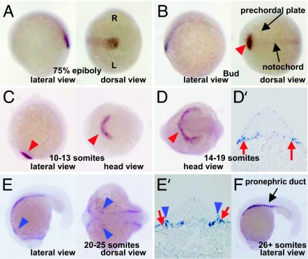
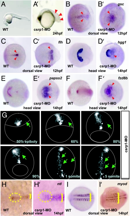
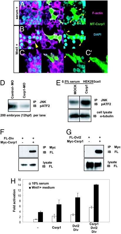
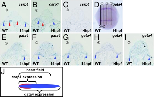
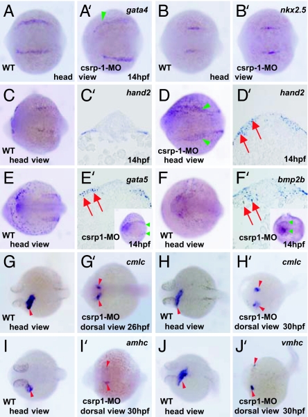
Zebrafish Csrp1 is a member of the cysteine- and glycine-rich protein (CSRP) family and is expressed in the mesendoderm and its derivatives. Csrp1 interacts with Dishevelled 2 (Dvl2) and Diversin (Div), which control cell morphology and other dynamic cell behaviors via the noncanonical Wnt and JNK pathways. When csrp1 message is knocked down, abnormal convergent extension cell movement is induced, resulting in severe deformities in midline structures. In addition, cardiac bifida is induced as a consequence of defects in cardiac mesoderm cell migration. Our data highlight Csrp1 as a key molecule of the noncanonical Wnt pathway, which orchestrates cell behaviors during dynamic morphogenetic movements of tissues and organs.


Conflict of interest statement

The authors declare no conflict of interest.

Figures

Fig. 1.
Fig. 1.
Expression patterns of zebrafish csrp1. (A) At 75% epiboly stage, csrp1 is expressed in the mesendoderm in the axis. R, right side of embryo; L, left side of embryo. (B) At the bud stage, expression is observed in the developing notochord and prechordal plate (arrowhead). (C) Expression of csrp1 becomes restricted in the prepolster region (arrowheads) at the 10- to 13-somite stage. (D) Expression is maintained in the polster at the 14- to 19-somite stage (arrowhead). (D′) Expression is also observed in the endoderm (arrows). (E and E′) Expression is detected in the endoderm beneath the cardiac mesoderm (red arrows), as well as in an anterior portion of the cardiac mesoderm (blue arrowheads) at the 14- to 19-somite stage. (F) At the 26-somite stage, csrp1 is expressed in the pronephric ducts.
Fig. 2.
Fig. 2.
Knockdown of csrp1 caused defects in gastrulation. (A and A′) Morphants of csrp1 had short bodies with compressed somites (arrowheads) compared with the WT (A). (B, B′, C, and C′) In the WT, marginal cells, which were marked by expression of gsc (B) and flh (C), accumulated at the margin. In the morphants, these cells did not accumulate, making large expression domains of gsc (B′) and flh (C′) at 12 hpf. Anterior migration of these cells was also impaired. (D–F and D′–F′) At 14 hpf, the prepolster region, marked by hgg1 (D) and papss2 (E), formed a U-shaped expression domain. In the morphants, this expression domain became deformed without a clear boundary (D′ and E′). (F and F′) Expression of fzd8b in the head mesenchyme was restricted in the WT at 14 hpf (F). This expression domain was expanded with dispersed signals in the morphants (F′). (G) Notochord cells were visualized by injection of pFlh-EGFP. In the morphants, marginal cells accumulated on the dorsal margin at 50% epiboly stage, but some cells were displaced without accumulation at the midline even at 60–80% epiboly stages (green arrows). At 90% epiboly stage, some mesen doderm cells (green arrows) localized beyond the territory of the notochord. Formation of the notochord was delayed and abnormal without elongation of cell shape. (H and H′) Morphology of the notochord was examined by ntl expression at 14 hpf. The faint but distinct accumulation of ntl-positive cells was observed in the anterior end of the notochord (circle in H). This group of cells was not observed in the morphants (circle in H′). The notochord of the morphants was wider than that of the WT (brackets in H and H′). These pictures were taken at the same magnification. (I and I′) Somites were visualized by expression of myod. In the morphants, thin and compressed somites were formed (bracket in I′) compared with the WT (bracket in I).
Fig. 3.
Fig. 3.
Csrp1 and the noncanonical Wnt/JNK signaling cascade. (A) Overexpression of Csrp1 in HEK293 cells induced formation of multiple cell protrusions (arrowheads) in the presence of 10% serum. In these protrusions, Csrp1 localized as shown by antibody staining. (B) When stimulated by Wnt1-conditioned medium, Csrp1-negative HEK293 cells elongated and formed small protrusions (arrowhead in Left). In contrast, Csrp1-expressing cells formed large protrusions where Csrp1 colocalized (arrowheads in Right). (C) Csrp1 accumulated in ruffling membrane formed at the edge of protrusions (arrowhead). A higher-magnification view is shown in C′. (D) Cell lysates prepared from zebrafish embryos were used for immunoprecipitation using an anti-JNK antibody. The amount of phosphorylated ATF2 in the precipitate was decreased in the csrp1 morphants, as revealed by an anti-phospho ATF2 antibody, compared with embryos injected with control MO. (E) Cell lysates were prepared from mock-transfected and Csrp1-expressing HEK293 cells. Phosphorylation of ATF2 was enhanced in the Csrp1-expressing cells. α-Tubulin was stained for a control. (F) From HEK293 cells expressing Flag-tagged Div (FL-Div) and Myc-tagged Csrp1 (Myc-Csrp1), Div was coprecipitated with Csrp1. (G) Immunoprecipitation assay using HEK293 cells. Flag-tagged Dvl2 (FL-Dvl2) and/or Myc-Csrp1 was introduced, and then Csrp1 was precipitated from lysates. In the precipitates, Dvl2 was detected. (H) A pAR-luciferase reporter and an expression vector for cJun were introduced in HEK293 cells along with Csrp1, Dvl2, and Div. This reporter is activated synergistically by Csrp1 and Dvl2/Div. This effect is further enhanced in the presence of Wnt1.
Fig. 4.
Fig. 4.
Expression of csrp1 in the cardiac mesoderm. Expression of csrp1 (A–C) and gata4 (E–I) was examined at 14 hpf in sections made at positions indicated in D. In an anterior region of the cardiac mesoderm that expresses gata4 (E), expression of csrp1 was observed in the mesoderm (blue arrowheads) and endoderm (red arrowheads) (A). (B and F) In a serial section, expression of csrp1 and gata4 was detected in the cardiac mesoderm (blue arrowheads). In this section, csrp1 is also expressed in the underlying endoderm (red arrowhead, B). (C and G) Expression of csrp1 was not detected in the next caudal sections, although gata4 expression was detected. (H and I) In a more posterior section, gata4 was still expressed. (J) A schematic illustration of the csrp1-positive and the gata4-positive domains in the heart field.
Fig. 5.
Fig. 5.
Cardiac bifida in the csrp1 morphants. (A–D and A′–D′) Expression of cardiac markers, such as gata4 or nkx2.5, was not affected in the csrp1 morphants at 14 hpf (A′ and B′, respectively), compared with the WT (A and B). In some cases, expression domains of these genes became faint and muddled (green arrowhead in A′). Note that the expression domain of gata4 shifted laterally, leaving a wider space in the middle. (C, C′, D, and D′) hand2, which is expressed in the cardiac mesoderm of the WT (C), was expressed in wider domains in the morphants (green arrowheads in D). This expansion was confirmed in sections (red arrows in D′) compared with the WT (C′). (E, E′, F, and F′) Expression of endodermal markers, such as gata5 (E and E′) and bmp2b (F and F′) was examined. Both genes were expressed in the endoderm beneath the cardiac mesoderm in the WT (E and F). In the morphants, expression of these genes became expanded (red arrows in sections and green arrowheads in whole mount, E′ and F′). (G and H) cmlc is normally expressed in the heart tube at 26 hpf (G) and 30 hpf (H). (G′ and H′) Cardiac mesoderm cells did not fuse to form a heart tube in the csrp1 morphants at 26 hpf (G′) and 30 hpf (H′). (I, I′, J, and J′) Likewise, as revealed by expression of amhc (I and I′) and vmhc (J and J′) at 30 hpf, cardiac mesoderm cells were kept separated.

Similar articles

Cited by

References

    1. Driever W. Curr Opin Genet Dev. 1995;5:610–618. - PubMed
    1. Myers DC, Sepich DS, Solnica-Krezel L. Trends Genet. 2002;18:447–455. - PubMed
    1. Heisenberg CP, Tada M, Rauch GJ, Saude L, Concha ML, Geisler R, Stemple DL, Smith JC, Wilson SW. Nature. 2000;405:76–81. - PubMed
    1. Kilian B, Mansukoski H, Barbosa FC, Ulrich F, Tada M, Heisenberg CP. Mech Dev. 2003;120:467–476. - PubMed
    1. Rauch GJ, Hammerschmidt M, Blader P, Schauerte HE, Strahle U, Ingham PW, McMahon AP, Haffter P. Cold Spring Harbor Symp Quant Biol. 1997;62:227–234. - PubMed

Publication types

MeSH terms