. 2007 Jul 3;104(27):11316-21.
doi: 10.1073/pnas.0702724104. Epub 2007 Jun 25.

Zebrafish model for human long QT syndrome

Affiliations

Zebrafish model for human long QT syndrome

Rima Arnaout et al. Proc Natl Acad Sci U S A. .

Abstract

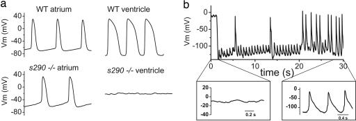
Long QT syndrome (LQTS) is a disorder of ventricular repolarization that predisposes affected individuals to lethal cardiac arrhythmias. To date, an appropriate animal model of inherited LQTS does not exist. The zebrafish is a powerful vertebrate model used to dissect molecular pathways of cardiovascular development and disease. Because fundamental electrical properties of the zebrafish heart are remarkably similar to those of the human heart, the zebrafish may be an appropriate model for studying human inherited arrhythmias. Here we describe the molecular, cellular, and electrophysiological basis of a zebrafish mutant characterized by ventricular asystole. Genetic mapping and direct sequencing identify the affected gene as kcnh2, which encodes the channel responsible for the rapidly activating delayed rectifier K(+) current (I(Kr)). We show that complete loss of functional I(Kr) in embryonic hearts leads to ventricular cell membrane depolarization, inability to generate action potentials (APs), and disrupted calcium release. A small hyperpolarizing current restores spontaneous APs, implying wild-type function of other ionic currents critical for AP generation. Heterozygous fish manifest overt cellular and electrocardiographic evidence for delayed ventricular repolarization. Our findings provide insight into the pathogenesis of homozygous kcnh2 mutations and expand the use of zebrafish mutants as a model system to study human arrhythmias.


Conflict of interest statement

The authors declare no conflict of interest.

Figures

Fig. 1.
Fig. 1.
Molecular analysis of two kcnh2 mutant alleles. (a–c) Wild-type and mutant embryos at 48 hpf, lateral oblique views, anterior to the left. Compared with wild type (WT) (a), s213 (b), and s290 (c), mutants exhibit a silent ventricle and, therefore, absent cardiac output and pronounced pericardial edema (red arrows). (d) The mutations map to linkage group 3. Flanking CA repeat markers and markers from selected BACs are shown, with the number of recombinants for the given number of meioses. The s213 and s290 mutations map to the potassium channel gene kcnh2. (e) Sequence analysis of kcnh2 in s213 mutants reveals a T–G transition at codon 462, resulting in an Ile–Arg substitution. For s290, a T–A transversion at codon 521 results in a Met–Lys substitution. (f) Schematic diagram showing the modular structure of Kcnh2. A Kcnh2 subunit consists of six transmembrane domains: the S1–S4 domains sense membrane potential, whereas the S5–S6 domains form the K-selective pore. Green and red stars represent the s213 and s290 mutation sites, respectively.
Fig. 2.
Fig. 2.
kcnh2 mutations cause complete loss of channel function. Representative ionic currents recorded from wild-type (WT), I462R, and M521K Kcnh2 heterologously expressed in Xenopus oocytes. Currents were elicited by 2-sec voltage steps between −80 and +40 mV applied from a holding potential of −80 mV. Deactivating tail currents were elicited by a 2-sec voltage step to −70 mV. Currents induced by I462R and M521K mutant Kcnh2 were no different from those seen in water-injected oocytes, indicating complete loss of channel function.
Fig. 3.
Fig. 3.
APs recorded from explanted embryonic zebrafish hearts. (a) (Upper) Representative spontaneous APs recorded from wild-type (WT) atrium (Left) and ventricle (Right) at 48 hpf. (Lower) Spontaneous APs recorded from kcnh2s290 mutant atrium (Left). Recordings of transmembrane voltage (Vm) from mutant ventricle (Right) revealed marked membrane depolarization and the absence of action potentials. (b) Vm recorded from kcnh2s290 mutant ventricle. Intracellular injection of hyperpolarizing current (−100 pA) caused membrane hyperpolarization and allowed for the generation of spontaneous APs.
Fig. 4.
Fig. 4.
Wild-type embryonic hearts in the Tg(cmlc2:gCaMP)s878 background exhibit atrial and ventricular conduction waves, whereas mutant hearts exhibit atrial, but no ventricular, conduction waves. SPIM videos of live 48 hpf hearts in the Tg(cmlc2:gCaMP)s878 background (see SI Movies 3 and 4) were processed to determine the fluorescence intensity of selected regions of the heart over time. Each selected region has a corresponding number plotted below. The dotted lines mark an arbitrary point in time to facilitate comparison across the different plots. (a) In a wild-type heart, fluorescence intensity varies with time in atrial and ventricular regions of the heart as the wave of depolarization propagates through the heart. This wave represents a wild-type heart rhythm. (b) In a kcnh2s290 mutant heart, fluorescence intensity varies in the atrium, but the ventricle maintains a constant, low-level intensity. The mutant heart lacks a ventricular conduction wave.
Fig. 5.
Fig. 5.
Heterozygous kcnh2 zebrafish manifest delayed ventricular repolarization. (a) APs recorded from 48 hpf heterozygous ventricle show increased AP duration compared with wild-type (see Results). (b) Representative electrocardiograms recorded from anesthetized, paralyzed wild-type and heterozygous adult zebrafish. Dashed line indicates duration of QT interval. QT corrected for heart rate (see Materials and Methods) was 396 and 467 msec for the exemplar wild-type and heterozygote, respectively.

References

    1. Sehnert AJS, Stainier DYR. Trends Genet. 2002;18:419–424. - PubMed
    1. Splawski I, Shen J, Timothy KW, Lehmann MH, Priori S, Robinson JL, Moss AJ, Schwartz PJ, Towbin JA, Vincent GM, et al. Circulation. 2000;102:1178–1185. - PubMed
    1. Hoorntje T, Alders M, van Tintelen P, van der Lip K, Sreeram N, van der Wal A, Mannens M, Wilde A. Circulation. 1999;100:1264–1267. - PubMed
    1. Schwartz PJ, Stramba-Badiale M. Prolonged Repolarization and Sudden Infant Death Syndrome. Philadelphia: Saunders; 2004.
    1. Babij P, Askew GR, Nieuwenhuijsen B, Su CM, Bridal TR, Jow B, Argentieri TM, Kulik J, DeGennaro LJ, Spinelli W, et al. Circ Res. 1998;83:668–678. - PubMed

Publication types

Substances