. 2007 Jul 3;104(27):11227-32.
doi: 10.1073/pnas.0703714104. Epub 2007 Jun 25.

Chymotrypsin C (caldecrin) promotes degradation of human cationic trypsin: identity with Rinderknecht's enzyme Y

Affiliations

Chymotrypsin C (caldecrin) promotes degradation of human cationic trypsin: identity with Rinderknecht's enzyme Y

Richárd Szmola et al. Proc Natl Acad Sci U S A. .

Abstract

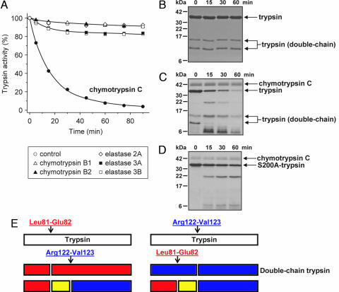
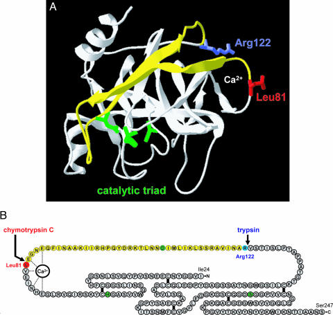
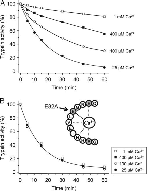
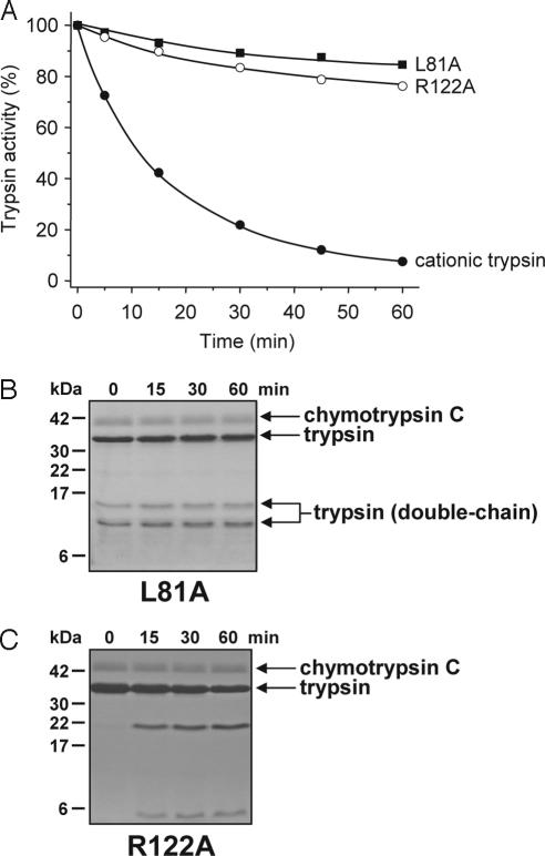
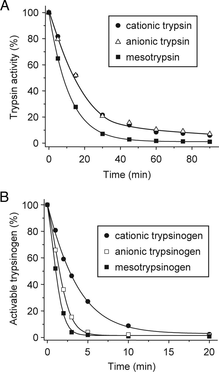
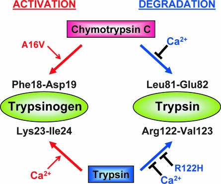
Digestive trypsins undergo proteolytic breakdown during their transit in the human alimentary tract, which has been assumed to occur through trypsin-mediated cleavages, termed autolysis. Autolysis was also postulated to play a protective role against pancreatitis by eliminating prematurely activated intrapancreatic trypsin. However, autolysis of human cationic trypsin is very slow in vitro, which is inconsistent with the documented intestinal trypsin degradation or a putative protective role. Here we report that degradation of human cationic trypsin is triggered by chymotrypsin C, which selectively cleaves the Leu(81)-Glu(82) peptide bond within the Ca(2+) binding loop. Further degradation and inactivation of cationic trypsin is then achieved through tryptic cleavage of the Arg(122)-Val(123) peptide bond. Consequently, mutation of either Leu(81) or Arg(122) blocks chymotrypsin C-mediated trypsin degradation. Calcium affords protection against chymotrypsin C-mediated cleavage, with complete stabilization observed at 1 mM concentration. Chymotrypsin C is highly specific in promoting trypsin degradation, because chymotrypsin B1, chymotrypsin B2, elastase 2A, elastase 3A, or elastase 3B are ineffective. Chymotrypsin C also rapidly degrades all three human trypsinogen isoforms and appears identical to enzyme Y, the enigmatic trypsinogen-degrading activity described by Heinrich Rinderknecht in 1988. Taken together with previous observations, the results identify chymotrypsin C as a key regulator of activation and degradation of cationic trypsin. Thus, in the high Ca(2+) environment of the duodenum, chymotrypsin C facilitates trypsinogen activation, whereas in the lower intestines, chymotrypsin C promotes trypsin degradation as a function of decreasing luminal Ca(2+) concentrations.


Conflict of interest statement

The authors declare no conflict of interest.

Figures

Fig. 1.
Fig. 1.
Degradation of human cationic trypsin by chymotrypsin C. (A) Cationic trypsin (2 μM) was incubated alone (control) or with 300 nM of the indicated proteases in 0.1 M Tris·HCl (pH 8.0) and 25 μM CaCl2 (final concentrations) in 100 μl of final volume. At the indicated times, 2-μl aliquots were withdrawn, and residual trypsin activity was measured and expressed as a percentage of the initial activity. (B–D) SDS/PAGE analysis of autolysis and chymotrypsin C-mediated degradation of cationic trypsin. Wild-type cationic trypsin (B and C) or S200A mutant cationic trypsin (D) were incubated at 2 μM concentration in the absence (B) or presence (C and D) of 300 nM chymotrypsin C in 0.1 M Tris·HCl (pH 8.0) and 25 μM CaCl2 (final concentrations). At the indicated times, 100-μl aliquots were precipitated with 10% trichloroacetic acid (final concentration) and electrophoresed on 15% minigels under reducing conditions, followed by Coomassie blue staining. Note that chymotrypsin C is glycosylated and runs as a fuzzy band. Double-chain trypsin is cleaved at the Arg122-Val123 peptide bond and runs as two bands on reducing gels; the upper band corresponds to the C-terminal chain (Val123-Ser247), and the lower band is the N-terminal chain (Ile24-Arg122). (E) Major proteolytic cleavage sites in cationic trypsin determined from N-terminal sequencing of the visible bands in C.
Fig. 2.
Fig. 2.
Structural determinants of chymotrypsin C-mediated trypsin degradation. (A) Ribbon diagram of human cationic trypsin [Protein Data Bank ID code 1TRN; chain B of the crystallographic dimer shown here (21)] with the Leu81 (red) and Arg122 (blue) side chains indicated. The calcium-binding loop is denoted by the Ca2+ symbol. The catalytic triad consisting of His63 (His57 in the conventional chymotrypsin numbering); Asp107 (chymotrypsin no. Asp102) and Ser200 (chymotrypsin no. Ser195) are shown in green. Note that Asp107 is located on the yellow peptide segment, which is released upon cleavage of the Leu81-Glu82 and Arg122-Val123 peptide bonds. See text for details. The image was rendered using DeepView/Swiss-PdbViewer version 3.7. (B) Primary structure of human cationic trypsin. Individual amino acids are represented by circles. The catalytic triad is highlighted in green, Leu81 is in red, and Arg122 is in blue. The five disulfide bridges and the interactions between the calcium ion and amino acids within the calcium binding loop are indicated. Note that both the Leu81-Glu82 and the Arg122-Val123 peptide bonds are located in a long peptide segment not stabilized by disulfide bonds. The yellow section corresponds to the yellow peptide in A.
Fig. 3.
Fig. 3.
Effect of calcium on the chymotrypsin C-mediated degradation of human cationic trypsin. Wild-type cationic trypsin (A) or the E82A mutant (B) were incubated at 2 μM concentration with 300 nM chymotrypsin C (final concentration) in 0.1 M Tris·HCl (pH 8.0) and the indicated CaCl2 concentrations. At the given times, residual trypsin activity was determined as described in Fig. 1A.
Fig. 4.
Fig. 4.
Mutations L81A and R122A stabilize cationic trypsin against chymotrypsin C-mediated degradation. Wild-type, L81A, and R122A cationic trypsins were incubated at 2 μM concentration with 300 nM chymotrypsin C in 0.1 M Tris·HCl (pH 8.0) and 25 μM CaCl2 (final concentrations). (A) At the indicated times, residual trypsin activity was measured as described in Fig. 1A. (B and C). Aliquots (100 μl) were precipitated with 10% trichloroacetic acid (final concentration) and analyzed by SDS/15% PAGE under reducing conditions.
Fig. 5.
Fig. 5.
Degradation of the three human trypsin and trypsinogen isoforms by chymotrypsin C. (A) Human cationic trypsin, anionic trypsin, and mesotrypsin were incubated at 2 μM concentration with 300 nM chymotrypsin C in 0.1 M Tris·HCl (pH 8.0) and 25 μM CaCl2 (final concentrations). At the indicated times, residual trypsin activity was measured as described in Fig. 1A. (B) Human cationic trypsinogen, anionic trypsinogen, and mesotrypsinogen (2 μM) were incubated with 300 nM chymotrypsin C in 0.1 M Tris·HCl (pH 8.0) and 25 μM CaCl2 (final concentrations). At the indicated times, 10-μl aliquots were withdrawn, CaCl2 was added to 10 mM final concentration and activable trypsinogen content was determined by incubating with 120 ng/ml human enteropeptidase for 15 min at room temperature and measuring trypsin activity. Activable trypsinogen was expressed as a percentage of the initial activable trypsinogen content.
Fig. 6.
Fig. 6.
Chymotrypsin C stimulates trypsin-mediated trypsinogen activation (autoactivation) and trypsin-mediated trypsin degradation (autolysis). In the presence of millimolar Ca2+ concentrations, the trypsin degradation pathway is blocked and only the trypsinogen activation pathway is operational. Both pathways are affected by certain hereditary pancreatitis-associated mutations. Mutation A16V stimulates the activation pathway (20), whereas mutation R122H inhibits the degradation pathway. See text for details.

References

    1. Guy O, Lombardo D, Bartelt DC, Amic J, Figarella C. Biochemistry. 1978;17:1669–1675. - PubMed
    1. Rinderknecht H, Renner IG, Carmack C. Gut. 1979;20:886–891. - PMC - PubMed
    1. Scheele G, Bartelt D, Bieger W. Gastroenterology. 1981;80:461–473. - PubMed
    1. Rinderknecht H, Renner IG, Abramson SB, Carmack C. Gastroenterology. 1984;86:681–692. - PubMed
    1. Rinderknecht H, Stace NH, Renner IG. Dig Dis Sci. 1985;30:65–71. - PubMed

Publication types

LinkOut - more resources