

Experimental validation of the Eclipse AAA algorithm
- PMID: 17592457
- PMCID: PMC5722411
- DOI: 10.1120/jacmp.v8i2.2350
Experimental validation of the Eclipse AAA algorithm
Abstract
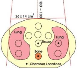
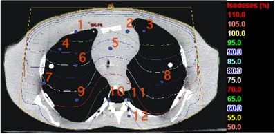
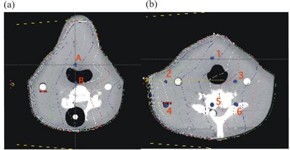
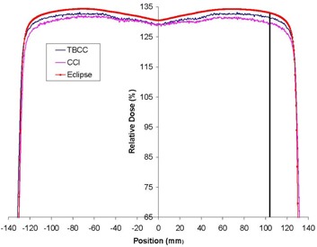
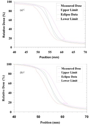
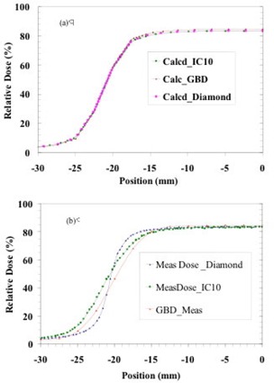
The present study evaluates the performance of a newly released photon-beam dose calculation algorithm that is incorporated into an established treatment planning system (TPS). We compared the analytical anisotropic algorithm (AAA) factory-commissioned with "golden beam data" for Varian linear accelerators with measurements performed at two institutions using 6-MV and 15-MV beams. The TG-53 evaluation regions and criteria were used to evaluate profiles measured in a water phantom for a wide variety of clinically relevant beam geometries. The total scatter factor (TSF) for each of these geometries was also measured and compared against the results from the AAA. At one institute, TLD measurements were performed at several points in the neck and thoracic regions of a Rando phantom; at the other institution, ion chamber measurements were performed in a CIRS inhomogeneous phantom. The phantoms were both imaged using computed tomography (CT), and the dose was calculated using the AAA at corresponding detector locations. Evaluation of measured relative dose profiles revealed that 97%, 99%, 97%, and 100% of points at one institute and 96%, 88%, 89%, and 100% of points at the other institution passed TG-53 evaluation criteria in the outer beam, penumbra, inner beam, and buildup regions respectively. Poorer results in the inner beam regions at one institute are attributed to the mismatch of the measured profiles at shallow depths with the "golden beam data." For validation of monitor unit (MU) calculations, the mean difference between measured and calculated TSFs was less than 0.5%; test cases involving physical wedges had, in general, differences of more than 1%. The mean difference between point measurements performed in inhomogeneous phantoms and Eclipse was 2.1% (5.3% maximum) and all differences were within TG-53 guidelines of 7%. By intent, the methods and evaluation techniques were similar to those in a previous investigation involving another convolution-superposition photon-beam dose calculation algorithm in another TPS, so that the current work permitted an independent comparison between the two algorithms for which results have been provided.
Figures
Similar articles
-
Testing of the analytical anisotropic algorithm for photon dose calculation.Med Phys. 2006 Nov;33(11):4130-48. doi: 10.1118/1.2358333. Med Phys. 2006. PMID: 17153392
-
Dosimetry of oblique tangential photon beams calculated by superposition/convolution algorithms: a Monte Carlo evaluation.J Appl Clin Med Phys. 2010 Nov 3;12(1):3424. doi: 10.1120/jacmp.v12i1.3424. J Appl Clin Med Phys. 2010. PMID: 21330989 Free PMC article.
-
Dosimetric Validation of Treatment Planning System for Volumetric Modulated Arc Therapy Using AAPM Medical Physics Practice Guideline 5.b.Asian Pac J Cancer Prev. 2024 May 1;25(5):1715-1723. doi: 10.31557/APJCP.2024.25.5.1715. Asian Pac J Cancer Prev. 2024. PMID: 38809644 Free PMC article.
-
Investigation of photon beam models in heterogeneous media of modern radiotherapy.Australas Phys Eng Sci Med. 2004 Jun;27(2):39-48. doi: 10.1007/BF03178375. Australas Phys Eng Sci Med. 2004. PMID: 15462585
-
Comparison of dose calculation algorithms in phantoms with lung equivalent heterogeneities under conditions of lateral electronic disequilibrium.Med Phys. 2004 Oct;31(10):2899-911. doi: 10.1118/1.1788932. Med Phys. 2004. PMID: 15543799
Cited by
-
Planning for mARC treatments with the Eclipse treatment planning system.J Appl Clin Med Phys. 2015 Mar 8;16(2):5351. doi: 10.1120/jacmp.v16i2.5351. J Appl Clin Med Phys. 2015. PMID: 26103202 Free PMC article.
-
Accuracy of out-of-field dose calculations by a commercial treatment planning system.Phys Med Biol. 2010 Dec 7;55(23):6999-7008. doi: 10.1088/0031-9155/55/23/S03. Epub 2010 Nov 12. Phys Med Biol. 2010. PMID: 21076191 Free PMC article.
-
Out-of-Field Dose Calculation by a Commercial Treatment Planning System and Comparison by Monte Carlo Simulation for Varian TrueBeam®.J Med Phys. 2019 Jul-Sep;44(3):156-175. doi: 10.4103/jmp.JMP_82_18. J Med Phys. 2019. PMID: 31576064 Free PMC article.
-
Accuracy Evaluation of EPL and ETAR Algorithms in the Treatment Planning Systems using CIRS Thorax Phantom.J Biomed Phys Eng. 2021 Aug 1;11(4):483-496. doi: 10.31661/jbpe.v0i0.1097. eCollection 2021 Aug. J Biomed Phys Eng. 2021. PMID: 34458196 Free PMC article.
-
Verification of IMRT dose calculations using AAA and PBC algorithms in dose buildup regions.J Appl Clin Med Phys. 2010 Aug 26;11(4):3351. doi: 10.1120/jacmp.v11i4.3351. J Appl Clin Med Phys. 2010. PMID: 21081894 Free PMC article.
References
-
- Cunningham J. Scatter‐air ratios. Phys Med Biol. 1972; 17 (1): 42–51. - PubMed
-
- Mackie TR, Scrimger J, Battista J. A convolution method of calculating dose for 15 MV X‐rays. Med Phys. 1985; 12 (2): 188–196. - PubMed
-
- Ahnesjö A, Saxner M, Trepp A. A pencil beam model photon dose calculation. Med Phys. 1992; 19 (2): 263–273. - PubMed
-
- Ulmer W, Pyyry J, Kaissl W. A 3D photon superposition/convolution algorithm and its foundation on results of Monte Carlo calculations. Phys Med Biol. 2005; 50 (8): 1767–1790. - PubMed
-
- Sievinen J, Ulmer W, Kaissl W. AAA photon dose calculation model in Eclipse. Palo Alto (CA): Varian Medical Systems; 2005: 1–18. [RAD #7170B]
Publication types
MeSH terms
LinkOut - more resources
Full Text Sources
Research Materials
Miscellaneous
