Review
. 2007 Aug;211(2):212-22.
doi: 10.1111/j.1469-7580.2007.00760.x. Epub 2007 Jun 25.

Distribution of neurons expressing tyrosine hydroxylase in the human cerebral cortex

Affiliations
Review

Distribution of neurons expressing tyrosine hydroxylase in the human cerebral cortex

Ruth Benavides-Piccione et al. J Anat. 2007 Aug.

Abstract

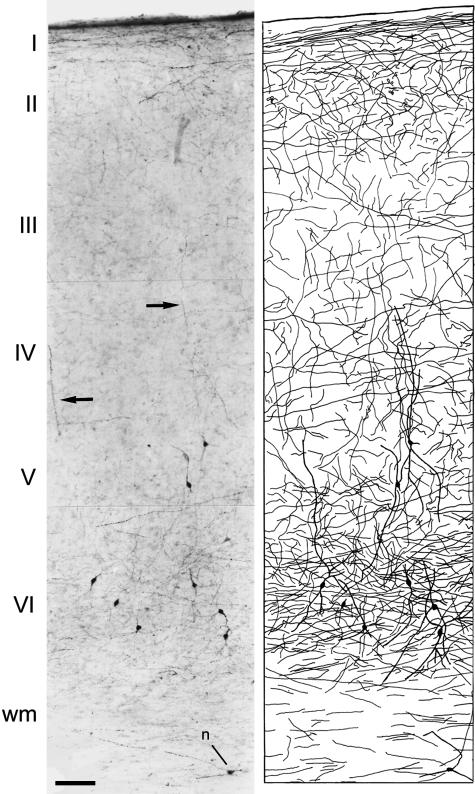
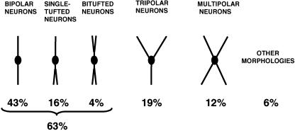
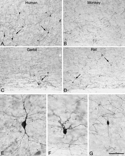
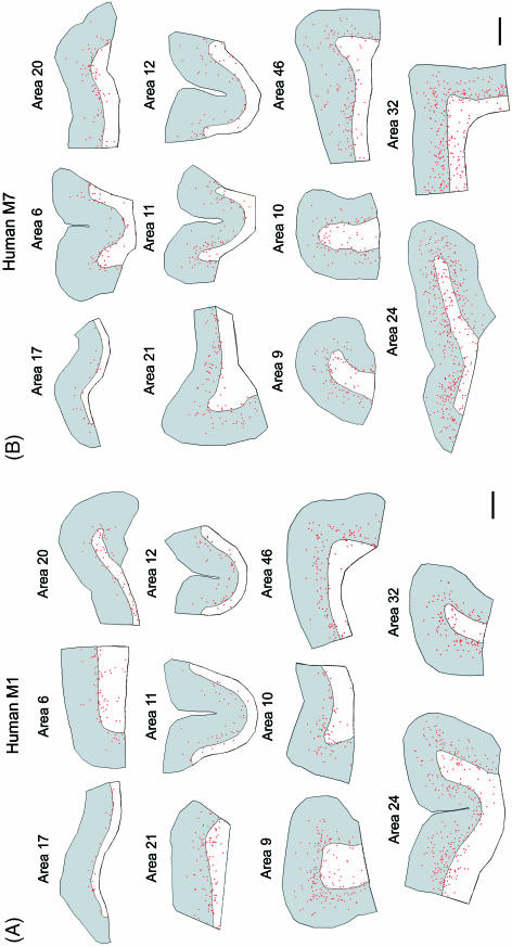
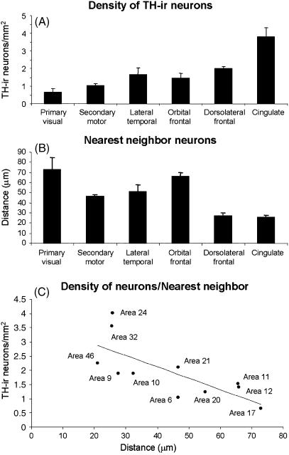
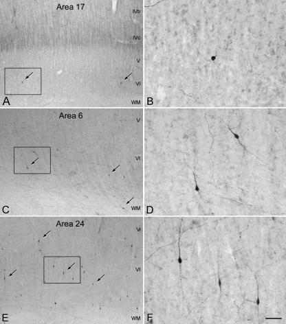
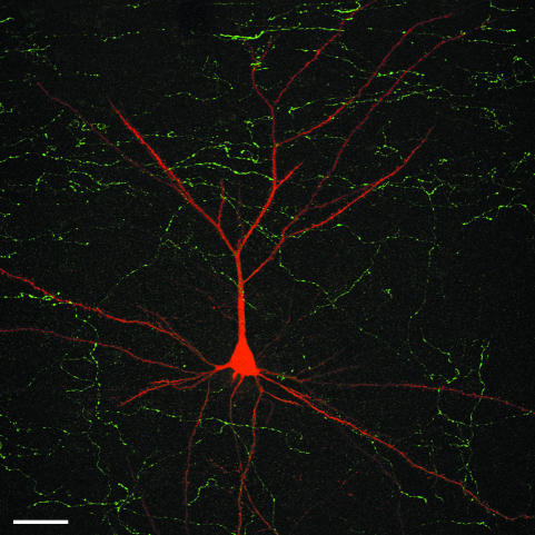
Since the very first detailed description of the different types of cortical interneurons by Cajal, the tremendous variation in the morphology, physiology and neurochemical properties of these cells has become apparent. However, it still remains unclear whether all types of interneurons are present in all cortical areas and species. Here we have focused on tyrosine hydroxylase (TH)-immunoreactive cortical interneurons, which although only present in certain species, are particularly abundant in the human neocortex. We argue that this type of interneuron is more widespread in the human neocortex than in any other species examined so far and that, therefore, it is probably involved in a larger variety of cortical circuits. In addition, notable regional variation can be seen in relation to these interneurons. These differences further emphasize the variability in the design of microcircuits between cortical areas and species, and they probably reflect an evolutionary adaptation of cortical circuits to particular functions.


Figures

Fig. 1
Fig. 1
Left: low-power photomicrograph from a biopsy of the human temporal cortex (Brodmann's area 20) showing TH immunostaining throughout layer I to the white matter. Note the labelling of neurons in layers V–VI. Arrows indicate vertically orientated fibres. n, a TH-ir neuron in the white matter. Right: Camera lucida drawing of the section on the left showing the laminar distribution of TH-ir neurons and fibres. Scale bar = 120 µm. Taken from Benavides-Piccione & DeFelipe (2003).
Fig. 2
Fig. 2
Schematic drawing showing the main morphological types of TH-ir neurons in layers V–VI of the human temporal cortex. Taken from Benavides-Piccione & DeFelipe (2003).
Fig. 3
Fig. 3
Low-power photomicrographs of TH immunostaining in the human temporal cortex (Brodmann's area 20) (A), macaque temporal cortex (area TE) (B), gerbil somatosensory cortex (C), and rat somatosensory cortex (D), extending from the lower part of layer V to the white matter. Some TH-ir neurons are indicated by arrows. E–G, high-power magnification of TH-ir neurons indicated (n) in A, C, and D, respectively. Scale bar = 160 µm for A–D and 40 µm for E–G. Taken from DeFelipe et al. (2006).
Fig. 4
Fig. 4
Camera lucida drawings showing the laminar distribution along layers I–VI (in grey) and underlying white matter (in white) of TH-ir neurons (red dots) in different cortical regions of the human M1 (A) and M7 (B). These regions correspond to the following Brodman's cytoarchitectonic subdivisions: primary visual area 17, secondary motor area 6, associative lateral temporal areas 20 and 21, associative orbital frontal areas 11 and 12, associative dorsolateral frontal areas 9, 10 and 46, and anterior limbic cingulate areas 24 and 32. Note the relative scarcity of TH-ir neurons in layers V–VI of area 17 and the high cell density in cingulate areas compared with other areas. Scale bar = 2 mm.
Fig. 5
Fig. 5
Plots showing the density of TH-ir neurons (A), nearest TH-ir cell neighbour analysis (B) and correlation analysis of these variables (C) from the primary visual cortex area 17, secondary motor area 6, associative lateral temporal cortex (areas 20 and 21), associative orbital prefrontal cortex (area 11 and 12), dorsolateral frontal cortex (area 9, 10 and 46) and anterior limbic cingulate cortex (areas 24 and 32). Data are expressed as the mean ± SEM. Statistical analysis showed that the differences in the TH-ir neuron density were significant (Kruskal–Wallis test H = 14.63, d.f. = 5, P < 0.05) between primary visual and limbic cingulate areas (post-hoc analyses, Dunn's multiple comparisons test, P < 0.05). Regarding TH-ir cell neighbour analysis, the distance between the closest pair of neurons in both dorsolateral frontal and cingulate cortex was significantly smaller than that in the orbital cortex (Kruskal–Wallis test H = 17.30, d.f. = 5, P < 0.005; post-hoc analyses, Dunn's multiple comparisons test, P < 0.05).
Fig. 6
Fig. 6
Photomicrographs showing TH-ir neurons (some indicated by arrows) and fibres in the deeper layers of areas 17 (A), 6 (C) and 24 (E) of the human neocortex. B, D and F are higher magnifications of the boxed areas in A, C and E, respectively. Note the differences in the number of TH-ir neurons between these three cortical areas. Scale bar = 190 µm in A, C and E, and 50 µm in B, D and F.
Fig. 7
Fig. 7
Confocal microscopy stack of 64 serial images showing an intracellularly injected pyramidal neuron (red) with Lucifer Yellow (LY) in the human temporal cortex, as well as the TH-ir fibres (green) in order to illustrate the relative location of TH-ir axons in the neuropil with respect to the pyramidal cell. The section was double-stained using a rabbit and mouse antiserum against LY and TH, respectively. The antibodies were visualized with biotinylated horse anti-mouse IgG and a mixture of Alexa fluor 594-conjugated goat anti-rabbit and streptavidin-conjugated Alexa fluor 488. Unpublished material from Benavides-Piccione et al. (2005). Scale bar = 40 µm.

References

    1. Akil M, Kolachana BS, Rothmond DA, Hyde TM, Weinberger DR, Kleinman JE. Catechol-O-methyltransferase genotype and dopamine regulation in the human brain. J Neurosci. 2003;23:2008–2013. - PMC - PubMed
    1. Ballesteros-Yáñez I, Muñoz A, Contreras J, Gonzalez J, Rodriguez-Veiga E, DeFelipe J. The double bouquet cell in the human cerebral cortex and a comparison with other mammals. J Comp Neurol. 2005;486:344–360. - PubMed
    1. Benavides-Piccione R, DeFelipe J. Different populations of tyrosine-hydroxylase-immunoreactive neurons defined by differential expression of nitric oxide synthase in the human temporal cortex. Cereb Cortex. 2003;13:297–307. - PubMed
    1. Benavides-Piccione R, Arellano J, DeFelipe J. Catecholamine innervation of pyramidal neurons in the human temporal cortex. Cereb Cortex. 2005;15:1584–1591. - PubMed
    1. Benes FM, Taylor JB, Cunningham MC. Convergence and plasticity of monoaminergic systems in the medial prefrontal cortex during the postnatal period: implications for the development of psychopathology. Cereb Cortex. 2000;10:1014–1027. - PubMed

MeSH terms

LinkOut - more resources