

Triterpenoids display single agent anti-tumor activity in a transgenic mouse model of chronic lymphocytic leukemia and small B cell lymphoma
- PMID: 17593960
- PMCID: PMC1891436
- DOI: 10.1371/journal.pone.0000559
Triterpenoids display single agent anti-tumor activity in a transgenic mouse model of chronic lymphocytic leukemia and small B cell lymphoma
Abstract
Background: The synthetic triterpenoid 2-Cyano-3,12-Dioxooleana-1,9-Dien-28-Oic Acid (CDDO) and derivatives display anti-tumor activity against a variety of cultured tumor cell lines and in mouse xenografts. In this report, we have studied the effects of CDDO and its imidazolide derivative (CDDO-Im) on chronic lymphocytic leukemia (CLL), using patients' CLL cells and a mouse model of CLL and small B cell lymphoma (SBL).
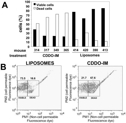
Principal findings: CDDO and CDDO-Im efficiently induced apoptosis of malignant human and mouse B-cells ex vivo, although CDDO-Im was over 10-fold more potent than CDDO. Treating mice with CLL/SBL with liposome-formulated CDDO or CDDO-Im resulted in significant reductions of B cells in blood, spleen and lung. CDDO-Im was shown to be more potent than CDDO, while treatment with empty liposomes had no impact on disease. CDDO-Im treatment initially resulted in an increase of circulating B cells, which correlates with a reduction in resident lymphocytes in spleen, and lungs, suggesting that CDDO-Im induces mobilization of tumor cells from lymphoid organs and infiltrated tissues into the circulation. Analysis of blood cells recovered from treated mice also showed that CDDO-Im is a potent inducer of tumor cells death in vivo. Furthermore, CDDO-Im efficiently eradicated mouse CLL/SBL cells but had little effect on the viability of normal B and T cells in vivo.
Significance: The presented data demonstrate that triterpenoids CDDO and CDDO-Im reduce leukemia and lymphoma burden in vivo in a transgenic mouse model of CLL/SBL, and support the clinical testing of CDDO-based synthetic triterpenoids in patients with CLL.
Conflict of interest statement
Figures


Similar articles
-
CDDO-Imidazolide inhibits growth and survival of c-Myc-induced mouse B cell and plasma cell neoplasms.Mol Cancer. 2006 Jun 7;5:22. doi: 10.1186/1476-4598-5-22. Mol Cancer. 2006. PMID: 16759389 Free PMC article.
-
Synthetic triterpenoids cooperate with tumor necrosis factor-related apoptosis-inducing ligand to induce apoptosis of breast cancer cells.Cancer Res. 2005 Jun 1;65(11):4799-808. doi: 10.1158/0008-5472.CAN-04-3319. Cancer Res. 2005. PMID: 15930300
-
The triterpenoid CDDO induces apoptosis in refractory CLL B cells.Blood. 2002 Oct 15;100(8):2965-72. doi: 10.1182/blood-2002-04-1174. Blood. 2002. PMID: 12351409
-
Preclinical evidences toward the use of triterpenoid CDDO-Me for solid cancer prevention and treatment.Mol Cancer. 2014 Feb 20;13:30. doi: 10.1186/1476-4598-13-30. Mol Cancer. 2014. PMID: 24552536 Free PMC article. Review.
-
CDDO and Its Role in Chronic Diseases.Adv Exp Med Biol. 2016;929:291-314. doi: 10.1007/978-3-319-41342-6_13. Adv Exp Med Biol. 2016. PMID: 27771930 Review.
Cited by
-
TNFR-associated factor 2 deficiency in B lymphocytes predisposes to chronic lymphocytic leukemia/small lymphocytic lymphoma in mice.J Immunol. 2012 Jul 15;189(2):1053-61. doi: 10.4049/jimmunol.1200814. Epub 2012 Jun 18. J Immunol. 2012. PMID: 22711886 Free PMC article.
-
Apoptosis inducers in chronic lymphocytic leukemia.Oncotarget. 2014 Jan 30;5(2):309-25. doi: 10.18632/oncotarget.1480. Oncotarget. 2014. PMID: 24525395 Free PMC article. Review.
-
The mitochondrial ATP-dependent Lon protease: a novel target in lymphoma death mediated by the synthetic triterpenoid CDDO and its derivatives.Blood. 2012 Apr 5;119(14):3321-9. doi: 10.1182/blood-2011-02-340075. Epub 2012 Feb 8. Blood. 2012. PMID: 22323447 Free PMC article.
-
Synthetic triterpenoids target the Arp2/3 complex and inhibit branched actin polymerization.J Biol Chem. 2010 Sep 3;285(36):27944-57. doi: 10.1074/jbc.M110.103036. Epub 2010 Jun 21. J Biol Chem. 2010. PMID: 20566646 Free PMC article.
-
The Traf2DNxBCL2-tg Mouse Model of Chronic Lymphocytic Leukemia/Small Lymphocytic Lymphoma Recapitulates the Biased IGHV Gene Usage, Stereotypy, and Antigen-Specific HCDR3 Selection of Its Human Counterpart.Front Immunol. 2021 Apr 12;12:627602. doi: 10.3389/fimmu.2021.627602. eCollection 2021. Front Immunol. 2021. PMID: 33912159 Free PMC article.
References
-
- Kipps T. Chronic lymphocytic leukemia. Curr Opin Hematol. 1997;4:268–276. - PubMed
-
- Reed JC, Kitada S. Apoptosis dysregulation in chronic lymphocytic leukemia. In: Cheson BD, editor. Chronic Lymphoid Leukemias. Second Edition ed. New York: Marcel Dekker, Inc; 2001. pp. 111–126.
-
- Elter T, Hallek M, Engert A. Fludarabine in chronic lymphocytic leukaemia. Expert Opin Pharmacother. 2006;7:1641–1651. - PubMed
-
- Kay NE. Purine analogue-based chemotherapy regimens for patients with previously untreated B-chronic lymphocytic leukemia. Semin Hematol. 2006;43:S50–54. - PubMed
-
- Kitada S, Pedersen IM, Schimmer A, Reed JC. Dysregulation of apoptosis genes in hematopeietic malignancies. Oncogene. 2002;21:3459–3474. - PubMed
Publication types
MeSH terms
Substances
Grants and funding
LinkOut - more resources
Full Text Sources
Other Literature Sources
Molecular Biology Databases
