. 2007 Jun 27;2(6):e571.
doi: 10.1371/journal.pone.0000571.

A highly sensitive assay for monitoring the secretory pathway and ER stress

Affiliations

A highly sensitive assay for monitoring the secretory pathway and ER stress

Christian E Badr et al. PLoS One. .

Abstract

Background: The secretory pathway is a critical index of the capacity of cells to incorporate proteins into cellular membranes and secrete proteins into the extracellular space. Importantly it is disrupted in response to stress to the endoplasmic reticulum that can be induced by a variety of factors, including expression of mutant proteins and physiologic stress. Activation of the ER stress response is critical in the etiology of a number of diseases, such as diabetes and neurodegeneration, as well as cancer. We have developed a highly sensitive assay to monitor processing of proteins through the secretory pathway and endoplasmic reticulum (ER) stress in real-time based on the naturally secreted Gaussia luciferase (Gluc).

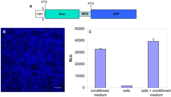
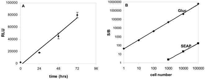
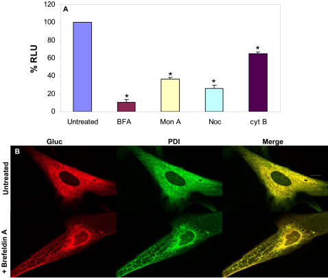
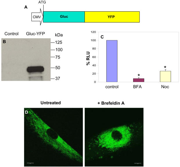
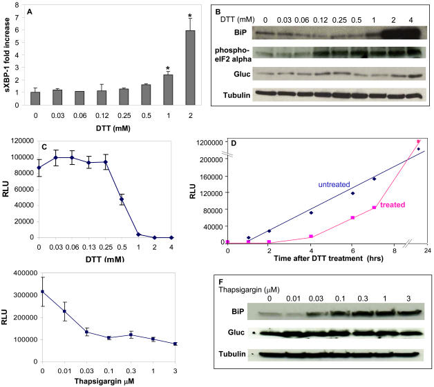
Methodology/principle findings: An expression cassette for Gluc was delivered to cells, and its secretion was monitored by measuring luciferase activity in the conditioned medium. Gluc secretion was decreased down to 90% when these cells were treated with drugs that interfere with the secretory pathway at different steps. Fusing Gluc to a fluorescent protein allowed quantitation and visualization of the secretory pathway in real-time. Expression of this reporter protein did not itself elicit an ER stress response in cells; however, Gluc proved very sensitive at sensing this type of stress, which is associated with a temporary decrease in processing of proteins through the secretory pathway. The Gluc secretion assay was over 20,000-fold more sensitive as compared to the secreted alkaline phosphatase (SEAP), a well established assay for monitoring of protein processing and ER stress in mammalian cells.

Conclusions/significance: The Gluc assay provides a fast, quantitative and sensitive technique to monitor the secretory pathway and ER stress and its compatibility with high throughput screening will allow discovery of drugs for treatment of conditions in which the ER stress is generally induced.


Conflict of interest statement

Competing Interests: The authors have declared that no competing interests exist.

Figures

Figure 1
Figure 1. Gluc as a reporter in mammalian cells.
(A) Schematic representation of the expression cassettes for Gluc-IRES-CFP cloned in the CSCW lentivirus vector. (B) High infection rate of cells with lentivirus vectors (M.O.I. = 30) as monitored by cerulean fluorescence. Scale bar, 100 µm. (C) Levels of Gluc activity in cells vs. medium vs. cells+medium. 293T cells were infected with the lentivirus vector carrying the expression cassette for Gluc-IRES-CFP and 20,000 cells were plated in wells of a 96-well plate. 48 hrs post-infection, new medium was added to the wells and Gluc activity was measured 24 hrs later in conditioned medium, viable washed cells or cells+conditioned medium after adding 2.5 µM coelenterazine.
Figure 2
Figure 2. Linearity and sensitivity of the Gluc assay.
(A) 293T cells were infected with the lentivirus vector carrying the expression cassette for Gluc-IRES-CFP and 20,000 cells were plated in wells of 96-well plate. Gluc activity was monitored over time in 10 µL of conditioned medium after addition of 20 µM coelenterazine. The release of Gluc to the conditioned medium is linear with time. (B) 293T cells were co-transfected with pHGC-Fluc and either pHGC-Gluc or pSEAP expression plasmids. Gluc and SEAP activities were assayed in conditioned medium 24 hrs later. The signal over background ratio (S/B) values, normalized to the intracellular levels of Fluc, are plotted against the number of cells. The Gluc assay can detect a single cell with S/B of 40 whereas the SEAP assay requires 20,000 cells to get similar S/B under similar assay conditions. The mean ± S.E.M. is presented on the graphs (n = 3).
Figure 3
Figure 3. Gluc as a reporter to monitor secretory pathway.
293T cells were infected with the lentivirus vector expressing Gluc and were plated in wells of 96-well plate. Cells were treated for 24 hrs with different drugs which interfere with the secretory pathway. (A) Cell-free conditioned media were assayed for Gluc activity which showed that Gluc secretion is decreased upon blocking the secretory pathway. *p≤0.01 as predicted by student T-test. (B) Immunocytochemistry on cells expressing Gluc with and without treatment with BFA showing that Gluc (cy3, red) co-localizes with the ER marker PDI (Alexa488, green). Scale bar, 10 µm.
Figure 4
Figure 4. Gluc-YFP fusion to visualize secretory pathway in real-time.
(A) Schematic representation of the Gluc-YFP fusion cloned in the lentivirus vector. (B) Uninfected 293T cells or cells infected with a lentivirus vector expressing Gluc-YFP fusion were lysed and analyzed by western blotting with anti-Gluc antibody. (C) Cells expressing Gluc-YFP fusion were treated with BFA or nocodazole and their conditioned medium were assayed for Gluc activity 24 hrs later. *p≤0.01 as predicted by student T-test. (D) Fluorescence microscopy of a single live cell expressing Gluc-YFP and either untreated or treated with BFA showing that this fusion is trapped in the ER upon BFA treatment. Scale bar, 10 µm.
Figure 5
Figure 5. Monitoring of ER stress with Gluc.
293T cells expressing Gluc were subjected to no or different concentrations of DTT (A–D) or thapsigargin (E–F) to induce ER stress. (A) Real-time RT-PCR for spliced XBP-1 mRNA which increased in response to >1 mM DTT 4 hrs after treatment. Fold induction is calculated with respect to the control non-infected/non-treated cells. (B&F) Western blot analysis showing upregulation of BiP and/or phosphorylated eIF2alpha levels in response to ER stress 24 hrs after treatment. Blot is also probed with Gluc antibody as well as beta-tubulin antibody for equal loading. (C&E) Conditioned medium assayed for Gluc activity 4 hrs after DTT or thapsigargin treatment showing that Gluc secretion is decreased in response to ER stress. (D) Untreated or treated cells with 1 mM DTT were monitored overtime for the level of Gluc secretion by assaying an aliquot of the conditioned medium for Gluc bioluminescence. The mean ± S.E.M. is presented on the graphs (n = 3), with *p≤0.01 as calculated by the student's t-Test.

References

    1. Ellgaard L, Helenius A. Quality control in the endoplasmic reticulum. Nat Rev Mol Cell Biol. 2003;4:181–191. - PubMed
    1. Voeltz GK, Rolls MM, Rapoport TA. Structural organization of the endoplasmic reticulum. EMBO Rep. 2002;3:994–950. - PMC - PubMed
    1. Stevens FJ, Argon Y. Pathogenic light chains and the B-cell repertoire. Immunol Today. 1999;20:451–457. - PubMed
    1. Tsai B, Ye Y, Rapoport TA. Retro-translocation of proteins from the endoplasmic reticulum into the cytosol. Nat Rev Mol Cell Biol. 2002;3:246–255. - PubMed
    1. Zhang K, Kaufman RJ. The unfolded protein response: a stress signaling pathway critical for health and disease. Neurology. 2006;66:S102–109. - PubMed

Publication types