. 2007 Jun 26:7:42.
doi: 10.1186/1472-6807-7-42.

X-ray structures of Na-GST-1 and Na-GST-2 two glutathione S-transferase from the human hookworm Necator americanus

Affiliations

X-ray structures of Na-GST-1 and Na-GST-2 two glutathione S-transferase from the human hookworm Necator americanus

Oluwatoyin A Asojo et al. BMC Struct Biol. .

Abstract

Background: Human hookworm infection is a major cause of anemia and malnutrition of adults and children in the developing world. As part of on-going efforts to control hookworm infection, The Human Hookworm Vaccine Initiative has identified candidate vaccine antigens from the infective L3 larval stages and adult stages of the parasite. Adult stage antigens include the cytosolic glutathione-S-transferases (GSTs). Nematode GSTs facilitate the inactivation and degradation of a variety of electrophilic substrates (drugs) via the nucleophilic addition of reduced glutathione. Parasite GSTs also play significant roles in multi-drug resistance and the modulation of host-immune defense mechanisms.

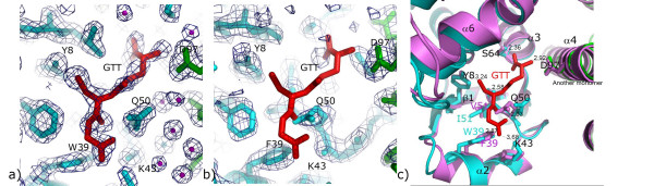
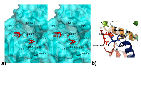
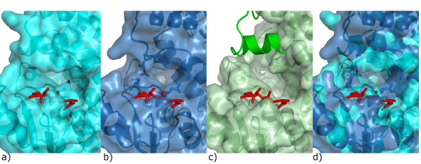
Results: The crystal structures of Na-GST-1 and Na-GST-2, two major GSTs from Necator americanus the main human hookworm parasite, have been solved at the resolution limits of 2.4 A and 1.9 A respectively. The structure of Na-GST-1 was refined to R-factor 18.9% (R-free 28.3%) while that of Na-GST-2 was refined to R-factor 17.1% (R-free 21.7%). Glutathione usurped during the fermentation process in bound in the glutathione binding site (G-site) of each monomer of Na-GST-2. Na-GST-1 is uncomplexed and its G-site is abrogated by Gln 50. These first structures of human hookworm parasite GSTs could aid the design of novel hookworm drugs.

Conclusion: The 3-dimensional structures of Na-GST-1 and Na-GST-2 show two views of human hookworm GSTs. While the GST-complex structure of Na-GST-2 reveals a typical GST G-site that of Na-GST-1 suggests that there is some conformational flexibility required in order to bind the substrate GST. In addition, the overall binding cavities for both are larger, more open, as well as more accessible to diverse ligands than those of GSTs from organisms that have other major detoxifying mechanisms. The results from this study could aid in the design of novel drugs and vaccine antigens.


Figures

Figure 1
Figure 1
Structural alignment of the a) four Na-GST-1 molecules and b) eight Na-GST-2 molecules in the crystallographic asymmetric unit, colored in rainbow from blue (N-termini) to red (C-termini). Side-chains in the regions of greatest variation are shown as sticks.
Figure 2
Figure 2
Ribbon representation of Na-GST-2 dimer reveals a typical GST dimer.
Figure 3
Figure 3
Sequence and structural alignment of Nu class GSTs with a Sigma Class GST (HsGST, human GST or hematopoietic prostagladin D synthase [37]) and other parasite GSTs (SjGST, Schistosoma japonica [43], Ascaris suum (As-GST-1) [42]. (a) The alignment reveals that firstly N-terminal alpha beta domain is more conserved than the C-terminal alpha domain. Furthermore, Na-GST-1 has higher sequence identity with HpolGST than Na-GST-2 and the lowest similarity is with the HsGST. This figure was generated with ESPript [55, 56]. (b) Structural alignment of monomers of Nu class GSTs (Na-GST-1, magenta; Na-GST-2, gold; HpolGST, green) with a sigma class GST (HsGST, cyan).
Figure 4
Figure 4
Comparison of GST dimers. a) Superposition of GST dimers reveals that they are very similar, however, Nu class (Na-GST-1, magenta; Na-GST-2, gold; HpolGST, green) have a more accessible binding cavity than sigma class (HsGST, cyan). The path to the binding cavity is indicated by the red arrow. The surface plots of Nu class GSTs b) HpolGST c) Na-GST-1 d) Na-GST-2 reveal larger access way to binding cavity than e) sigma class GST (HsGST).
Figure 5
Figure 5
G-site features. a) G-site of Na-GST-2 shows unambiguous electron density for glutathione (GTT) in 2Fo-Fc maps contoured at 1 sigma. b) No such density is visible in the G-site of Na-GST-1. c) Alignment of G-sites of Na-GST-2 and Na-GST-1. GTT is modeled from Na-GST-2 structure. The monomers of Na-GST-2 dimer are colored in cyan and green, while Na-GST-1 is colored in violet. Trp39 forms a hydrogen bond with glutathione in Na-GST-2 which is replaced with Phe39 in Na-GST-1. Gln50 is conserved in both Na-GST-2 and Na-GST-1, but the side chain is flipped such that glutathione cannot fit in Na-GST-1 G-site. The catalytic Tyr8 maintains its conformation in both structures. Polar interactions and distances are also shown.
Figure 6
Figure 6
H-site features. a) Stereo representation of the H-site of Na-GST-2, residues that form the cleft are shown as sticks. b) The C-terminal loop of Na-GST-2 is stabilized by a network of hydrogen bonds, residues involved in these interactions are shown in as sticks, while the bonds are indicated by dashed lines and distances are shown. Residues are colored in rainbow representation from N-terminal to C-terminal (Blue-Green-Yellow-Orange-Red). Two ethylene glycol molecules (EDO, in cyan) from the cryo-protecting liquor are visible in the surface of the H-site.
Figure 7
Figure 7
Nu class GSTs {a) Na-GST-2, c) HpolGST} have larger binding cavity than sigma class GST {b) HsGST}. d) Overlay of the cavities reveals the considerable reduction in the active site size between sigma class (blue) and nu class (cyan). The structure of HpolGST is missing a loop in close proximity to the binding cavity and we modeled it as cartoon from the Na-GST-2 structure. The glutathione in the G-site is shown as red stick model.

References

    1. Hotez PJ, Bethony J, Bottazzi ME, Brooker S, Buss P. Hookworm: "the great infection of mankind". PLoS Med. 2005;2:e67. doi: 10.1371/journal.pmed.0020067. - DOI - PMC - PubMed
    1. Bethony J, Brooker S, Albonico M, Geiger SM, Loukas A, Diemert D, Hotez PJ. Soil-transmitted helminth infections: ascariasis, trichuriasis, and hookworm. Lancet. 2006;367:1521–1532. doi: 10.1016/S0140-6736(06)68653-4. - DOI - PubMed
    1. Crompton DW. The public health importance of hookworm disease. Parasitology. 2000;121:S39–50. doi: 10.1017/S0031182000006454. - DOI - PubMed
    1. Hotez PJ, Brooker S, Bethony JM, Bottazzi ME, Loukas A, Xiao S. Hookworm infection. N Engl J Med. 2004;351:799–807. doi: 10.1056/NEJMra032492. - DOI - PubMed
    1. Bleakley H. Disease and Development: Evidence from the American South. Journal of the European Economic Association. 2003;1:376–386. doi: 10.1162/154247603322391017. - DOI

Publication types

MeSH terms

Substances