. 2007 Aug 29;362(1484):1357-68.
doi: 10.1098/rstb.2007.2121.

Biomimetic approach to cardiac tissue engineering

Affiliations

Biomimetic approach to cardiac tissue engineering

M Radisic et al. Philos Trans R Soc Lond B Biol Sci. .

Abstract

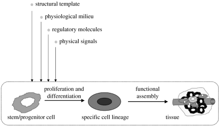
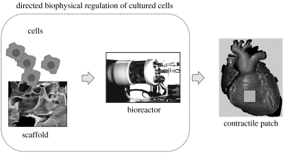
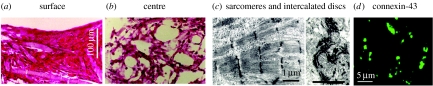
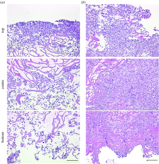
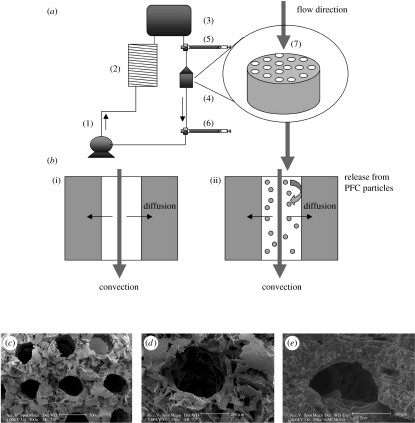
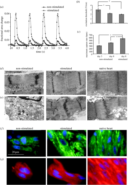
Here, we review an approach to tissue engineering of functional myocardium that is biomimetic in nature, as it involves the use of culture systems designed to recapitulate some aspects of the actual in vivo environment. To mimic the capillary network, subpopulations of neonatal rat heart cells were cultured on a highly porous elastomer scaffold with a parallel array of channels perfused with culture medium. To mimic oxygen supply by haemoglobin, the culture medium was supplemented with a perfluorocarbon (PFC) emulsion. Constructs cultivated in the presence of PFC contained higher amounts of DNA and cardiac markers and had significantly better contractile properties than control constructs cultured without PFC. To induce synchronous contractions of cultured constructs, electrical signals mimicking those in native heart were applied. Over only 8 days of cultivation, electrical stimulation induced cell alignment and coupling, markedly increased the amplitude of synchronous construct contractions and resulted in a remarkable level of ultrastructural organization. The biomimetic approach is discussed in the overall context of cardiac tissue engineering, and the possibility to engineer functional human cardiac grafts based on human stem cells.


Figures

Figure 1
Figure 1
Developmental paradigm. Tissue development and remodelling, in vivo and in vitro, involve the proliferation and differentiation of stem/progenitor cells and their subsequent assembly into a tissue structure. Cell function and the progression of tissue assembly depend on (i) the availability of a scaffold for cell attachment and tissue formation, (ii) the maintenance of physiological conditions in cell/tissue environment, (iii) the supply of nutrients, oxygen, metabolites and growth factors, and (iv) the presence of physical regulatory factors (adapted from Radisic et al. 2005b).
Figure 2
Figure 2
Tissue engineering paradigm. The regulatory factors of cell differentiation and tissue assembly shown in figure 1 can be used in vitro to engineer functional tissues by an integrated use of isolated cells, biomaterial scaffolds and bioreactors. The cells themselves (either differentiated or progenitor/stem cells seeded onto a scaffold and cultured in a bioreactor) carry out the process of tissue formation in response to regulatory signals. The scaffold provides a structural, mechanical and logistic template for cell attachment and tissue formation. The bioreactor provides the environmental conditions and regulatory signals (biochemical and physical) that induce, enhance or at least support the development of functional tissue constructs (adapted from Radisic et al. 2005b).
Figure 3
Figure 3
Cardiac constructs engineered in a conventional culture system. Constructs seeded and cultured with medium flow at construct surfaces formed (a) approximately 100 μm thick peripheral region (b) around an acellular interior. Cells in the peripheral region were electrically connected via gap junctions (d), immunostain for connexin-43 (c) exhibited cardiac-specific ultrastructural features, and propagated electrical signals over a distance of 5 mm (as recorded by a linear array of electrodes; Papadaki et al. 2001) (adapted from Carrier et al. 1999).
Figure 4
Figure 4
Effects of perfusion during seeding and cultivation on cardiac cell distribution. Cross-sections of constructs inoculated with 12 million cells and then transferred for a period of 4.5 h either into (a) dishes (25 r.p.m.) or into (b) perfused cartridges (1.5 ml min−1). The top, centre and bottom areas of a 650 μm wide strip extending from one construct surface to the other are shown. Scale bar, 100 μm (adapted from Radisic et al. 2003).
Figure 5
Figure 5
Oxygen supply: medium perfusion, channelled scaffolds and oxygen carriers. (a) Perfusion loop. Channelled biorubber scaffolds (7) preconditioned with cardiac fibroblasts were seeded with CMs and placed into perfusion cartridges (4) between two debubbling syringes (5,6). Medium flow (0.1 ml min−1) was provided by a multi-channel peristaltic pump (1) and gas exchange was provided by a coil of thin silicone tubing (2). Loops were placed in the 37oC/5% CO2 incubator vertically. (b) Modes of oxygen transport in the channelled construct perfused with culture medium include convection through the channel lumen and diffusion into the tissue space surrounding each channel. In regular culture medium ((i) control group), oxygen dissolved in the aqueous phase during gas exchange in the external loop is transported into the tissue phase and consumed by the cells. In culture medium supplemented with 10% PFC emulsion ((ii) PFC group), oxygen is replenished within the tissue construct by the release of oxygen from the PFC particles into the culture medium phase. Scanning electron micrographs of biorubber scaffold with (c) a parallel channel array and (d) of a single channel are shown at the beginning and (e) after 3 days of cultivation (adapted from Radisic et al. (2005a)).
Figure 6
Figure 6
Effects of electrical stimulation on functional assembly of engineered cardiac constructs. (a) Contraction amplitude of constructs cultured for a total of 8 days, shown as a fractional change in the construct size. Electrical stimulation increased the amplitude of contractions by a factor of 7. (b) ET decreased and (c) MCR increased significantly both with time in culture and due to electrical stimulation. (*) denotes statistically significant differences (p<0.05; Tukey's post hoc test with one-way ANOVA, n=5–10 samples per group and time point). (d) The structure of sarcomeres and (e) gap junctions observed in micrographs of stimulated constructs after 8 days of cultivation were remarkably similar to neonatal rat ventricles and markedly better developed than in control (non-stimulated) constructs. Representative sections of constructs stained for (f) Cx-43 (green) and (g) β-MHC (red), cell nuclei are shown in blue (adapted from Radisic et al. (2004a)).

Similar articles

Cited by

References

    1. Akins R.E, Boyce R.A, Madonna M.L, Schroedl N.A, Gonda S.R, McLaughlin T.A, Hartzell C.R. Cardiac organogenesis in vitro: reestablishment of three-dimensional tissue architecture by dissociated neonatal rat ventricular cells. Tissue Eng. 1999;5:103–118. doi:10.1089/ten.1999.5.103 - DOI - PubMed
    1. Balsam L.B, Wagers A.J, Christensen J.L, Kofidis T, Weissman I.L, Robbins R.C. Haematopoietic stem cells adopt mature haematopoietic fates in ischaemic myocardium. Nature. 2004;428:668–673. doi:10.1038/nature02460 - DOI - PubMed
    1. Beltrami A.P, et al. Adult cardiac stem cells are multipotent and support myocardial regeneration. Cell. 2003;114:763–776. doi:10.1016/S0092-8674(03)00687-1 - DOI - PubMed
    1. Brilla C.G, Maisch B, Rupp H, Sunck R, Zhou G, Weber K.T. Pharmacological modulation of cardiac fibroblast function. Herz. 1995;20:127–135. - PubMed
    1. Burlew B.S, Weber K.T. Cardiac fibrosis as a cause of diastolic dysfunction. Herz. 2002;27:92–98. doi:10.1007/s00059-002-2354-y - DOI - PubMed

Publication types

LinkOut - more resources