

U bodies are cytoplasmic structures that contain uridine-rich small nuclear ribonucleoproteins and associate with P bodies
- PMID: 17595295
- PMCID: PMC1899408
- DOI: 10.1073/pnas.0704977104
U bodies are cytoplasmic structures that contain uridine-rich small nuclear ribonucleoproteins and associate with P bodies
Abstract
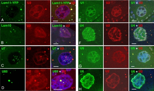
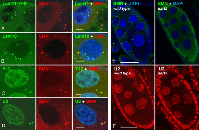
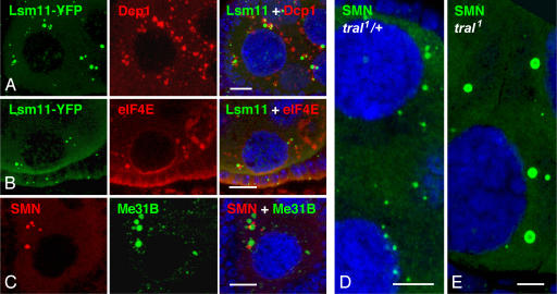
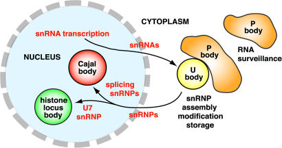
Uridine-rich small nuclear ribonucleoproteins (U snRNPs) are involved in key steps of pre-mRNA processing in the nucleus of eukaryotic cells. U snRNPs are enriched in the nucleus in discrete organelles that include speckles, Cajal bodies, and histone locus bodies. However, most U snRNPs are assembled in the cytoplasm, not in the nucleus. Despite extensive biochemical information, little is known about the spatial organization of U snRNPs in the cytoplasm. Here we show that U snRNPs in Drosophila are concentrated in discrete cytoplasmic structures, which we call U bodies, because they contain the major U snRNPs. In addition to snRNPs, U bodies contain essential snRNP assembly factors, suggesting that U bodies are sites for assembly or storage of snRNPs before their import into the nucleus. U bodies invariably associate with P bodies, which are involved in RNA surveillance and decay. Genetic disruption of P body components affects the organization of U bodies, suggesting that the two cytoplasmic bodies may cooperate in regulating aspects of snRNP metabolism. The identification of U bodies provides an opportunity to correlate specific biochemical steps of snRNP biogenesis with structural features of the cytoplasm.
Conflict of interest statement
The authors declare no conflict of interest.
Figures
Similar articles
-
Drosophila SMN complex proteins Gemin2, Gemin3, and Gemin5 are components of U bodies.Exp Cell Res. 2010 Aug 15;316(14):2354-64. doi: 10.1016/j.yexcr.2010.05.001. Epub 2010 May 7. Exp Cell Res. 2010. PMID: 20452345
-
A role for Cajal bodies in the final steps of U2 snRNP biogenesis.J Cell Sci. 2004 Sep 1;117(Pt 19):4423-33. doi: 10.1242/jcs.01308. Epub 2004 Aug 17. J Cell Sci. 2004. PMID: 15316075
-
The spinal muscular atrophy disease gene product, SMN: A link between snRNP biogenesis and the Cajal (coiled) body.J Cell Biol. 1999 Nov 15;147(4):715-28. doi: 10.1083/jcb.147.4.715. J Cell Biol. 1999. PMID: 10562276 Free PMC article.
-
Joining the dots: production, processing and targeting of U snRNP to nuclear bodies.Biochim Biophys Acta. 2008 Nov;1783(11):2137-44. doi: 10.1016/j.bbamcr.2008.07.025. Epub 2008 Aug 6. Biochim Biophys Acta. 2008. PMID: 18725249 Review.
-
Nuclear import of spliceosomal snRNPs.Can J Physiol Pharmacol. 2006 Mar-Apr;84(3-4):367-76. doi: 10.1139/y05-101. Can J Physiol Pharmacol. 2006. PMID: 16902583 Review.
Cited by
-
Identification of novel filament-forming proteins in Saccharomyces cerevisiae and Drosophila melanogaster.J Cell Biol. 2010 Aug 23;190(4):541-51. doi: 10.1083/jcb.201003001. Epub 2010 Aug 16. J Cell Biol. 2010. PMID: 20713603 Free PMC article.
-
Analysis of asymptomatic Drosophila models for ALS and SMA reveals convergent impact on functional protein complexes linked to neuro-muscular degeneration.BMC Genomics. 2023 Sep 27;24(1):576. doi: 10.1186/s12864-023-09562-4. BMC Genomics. 2023. PMID: 37759179 Free PMC article.
-
Components of the RNAi machinery that mediate long-distance chromosomal associations are dispensable for meiotic and early somatic homolog pairing in Drosophila melanogaster.Genetics. 2008 Nov;180(3):1355-65. doi: 10.1534/genetics.108.092650. Epub 2008 Sep 14. Genetics. 2008. PMID: 18791234 Free PMC article.
-
Spinal Muscular Atrophy: From Defective Chaperoning of snRNP Assembly to Neuromuscular Dysfunction.Front Mol Biosci. 2017 Jun 8;4:41. doi: 10.3389/fmolb.2017.00041. eCollection 2017. Front Mol Biosci. 2017. PMID: 28642865 Free PMC article. Review.
-
Survival motor neuron protein regulates stem cell division, proliferation, and differentiation in Drosophila.PLoS Genet. 2011 Apr;7(4):e1002030. doi: 10.1371/journal.pgen.1002030. Epub 2011 Apr 7. PLoS Genet. 2011. PMID: 21490958 Free PMC article.
References
-
- Steitz JA, Black L, Gerke V, Parker KA, Kramer A, Frendeway D, Keller W. In: Structure and Function of Major and Minor Small Nuclear Ribonucleoproteins. Birnstiel ML, editor. Berlin: Springer; 1998. pp. 115–154.
-
- Nilsen TW. BioEssays. 2003;25:1147–1149. - PubMed
-
- Tycowski KT, Kolev NG, Conrad NK, Fok V, Steitz JA. In: The RNA World. Gesteland RF, Cech TR, Atkins JF, editors. Cold Spring Harbor, NY: Cold Spring Harbor Lab Press; 2006. pp. 327–368.
-
- Marzluff WF. Curr Opin Cell Biol. 2005;17:274–280. - PubMed
Publication types
MeSH terms
Substances
Grants and funding
LinkOut - more resources
Full Text Sources
Other Literature Sources
Molecular Biology Databases
Miscellaneous
