. 2007 Sep;98(9):1491-7.
doi: 10.1111/j.1349-7006.2007.00534.x. Epub 2007 Jul 26.

Novel antiangiogenic pathway of thrombospondin-1 mediated by suppression of the cell cycle

Affiliations

Novel antiangiogenic pathway of thrombospondin-1 mediated by suppression of the cell cycle

Mai Yamauchi et al. Cancer Sci. 2007 Sep.

Abstract

We have recently reported that keratin 14-promoter-driven vascular endothelial growth factor (VEGF)-E(NZ-7) transgenic mice have a significant number of capillary vessels in subcutaneous tissue. However, these vessels are generated in a layer some distance from the epithelial basal cells that express VEGF-E(NZ-7), suggesting that one or more antiangiogenenic molecules may exist very near the basal cell layer. By screening keratinocyte-conditioned medium, we found that thrombospondin-1 (TSP-1) is produced from keratinocytes and suppresses human umbilical vein endothelial cells (HUVEC) growth as well as tubular formation in a HUVEC-fibroblast coculture system. Different to the known mechanism of CD36-dependent endothelial cell apoptosis, the HUVEC we used did not express CD36 at detectable levels, indicating a new mechanism for TSP-1-induced antiangiogenesis. We found that TSP-1 induces little apoptosis of endothelial cells but causes cell-cycle arrest, increasing the amounts of p21(CIP/WAF-1) and unphosphorylated retinoblastoma (Rb) in HUVEC. CD36-binding peptide in TSP-1 and CD36-neutralizing antibody did not block the TSP-1-induced cell-cycle arrest. Our results strongly suggest that TSP-1 utilizes a novel pathway for its antiangiogenic effect independent of CD36, and suppresses the cell cycle.


Figures
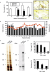
Figure 1
Figure 1
A major antiangiogenic factor secreted from keratinocytes is thrombospondin‐1 (TSP‐1). (A) Human keratinocyte (HK) conditioned medium (CM) inhibits tubular formation of human umbilical vein endothelial cells (HUVEC) in a HUVEC–fibroblast coculture system. The results in the left panel indicate tube length with or without HK CM. The right panel shows representative tubular structures with (50%) or without HK CM. (B) Fractions 9 and 10 from the gel chromatography inhibit the proliferation and tubular formation of HUVEC. (C) Silver‐stained sodium dodecylsulfate–polyacrylamide gel electrophoresis for concentrated HK CM and control medium. The TSP‐1‐specific band indicated with an asterisk at 180 kDa was detected by western blotting using anti‐TSP‐1 antibody (right panel). (D,E) TSP‐1 inhibits the tubular formation (D) and proliferation of HUVEC (E) in a dose‐dependent manner. Results represent the mean ± SD from several independent experiments.
Figure 2
Figure 2
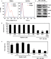
Thrombospondin‐1 (TSP‐1) inhibits angiogenesis in a CD36‐independent manner. (A) Expression of CD36 is undetectable on human umbilical vein endothelial cells (HUVEC) using flow cytometry. Peripheral blood mononuclear cells (PBMC): a positive control collected from human peripheral blood using Ficoll‐Paque plus. (B,C) A synthetic peptide (peptide A), which blocks CD36–TSP‐1 binding, does not inhibit the tubular formation and proliferation of HUVEC. A neutralizing antibody against TSP‐1 named Ab‐1, which binds to the CD36 recognition site on TSP‐1, and CD36 neutralizing antibody do not rescue the inhibition of HUVEC proliferation by TSP‐1. TSP‐1 and Ab‐1 (50 µg/mL) were preincubated for 1 h before use. Anti‐CD36 antibody (10 µg/mL) was added 1 h before the addition of TSP‐1. Results represent the mean ± SD of several independent experiments. (D) Phosphorylation of either p38 or JNK was not detected in HUVEC treated with TSP‐1 using western blotting. The HUVEC sample was obtained 30 min after addition of TSP‐1.
Figure 3
Figure 3
Thrombospondin‐1 (TSP‐1) induces growth arrest in human umbilical vein endothelial cells (HUVEC). (A) Ki‐67‐positive proliferating cells dramatically decreased in number among TSP‐1‐treated HUVEC. Representative results for the TSP‐1‐treated HUVEC are shown in the upper panels. Hoechst 33258 was used for nuclear staining. The graph below shows the percentage of Ki‐67‐positive cells. (B) TSP‐1 impairs S‐phase progression in HUVEC. Cell‐cycle analysis by flow cytometry was carried out using HUVEC treated with TSP‐1 (25 nM) for 72 h. The results indicate a quantified ratio of cells at each phase as the mean percentage ± SE from several independent experiments.
Figure 4
Figure 4
Upregulation of p21Cip1/WAF‐1 expression, phosphorylation of p53, and dephosphorylation of retinoblastoma (Rb) in human umbilical vein endothelial cells (HUVEC) treated with thrombospondin‐1 (TSP‐1). (A) HUVEC were treated with or without TSP‐1 (25 nM) at 37°C for 24 h. Cell lysates were used directly for sodium dodecylsulfate–polyacrylamide gel electrophoresis (SDS‐PAGE), and analyzed by western blotting using the specific antibodies indicated at the right of the figure. The graph below shows quantified results obtained using NIH image 1.6 software. (B) HUVEC were treated with or without TSP‐1 (25 nM) at 37°C for 15, 30 and 60 min. Cell lysates were separated by SDS‐PAGE, and analyzed by western blotting using the antibodies indicated at the right of the figure. Representative results are shown in the left panels. The graph shows quantified results obtained using NIH image 1.6 software.
Figure 5
Figure 5
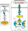
Schematic model of the effects of thrombospondin‐1 (TSP‐1) on endothelial cells dependent on or independent of CD36. TSP‐1 is reported to bind CD36, activate caspases through p38 and JNK, and induce apoptosis in endothelial cells expressing CD36 (left). However, our results suggest that TSP‐1 induces endothelial cell growth arrest mediated by an upregulation of p21CIP/WAF‐1 expression, phosphorylation of p53, and dephosphorylation of Rb in a CD36‐independent manner (right).

Similar articles

Cited by

References

    1. Folkman J. Tumor angiogenesis: therapeutic implications. N Eng J Med 1991; 285: 1182–6. - PubMed
    1. Hanahan D, Folkman J. Patterns and emerging mechanisms of the angiogenic switch during tumorigenesis. Cell 1996; 86: 353–64. - PubMed
    1. Risau W. Mechanisms of angiogenesis. Nature 1997; 386: 671–4. - PubMed
    1. Kiba A, Sagara H, Hara T, Shibuya M. VEGFR‐2‐specific ligand VEGF‐E induces non‐edematous hyper‐vascularization in mice. Biochem Biophys Res Commun 2003; 301: 371–7. - PubMed
    1. Folkman J. Endogenous angiogenesis inhibitors. APMIS 2004; 112: 496–507. - PubMed

Publication types

MeSH terms

LinkOut - more resources