. 2007 Sep;81(17):8891-904.
doi: 10.1128/JVI.00614-07. Epub 2007 Jun 27.

A rapid progressor-specific variant clone of simian immunodeficiency virus replicates efficiently in vivo only in the absence of immune responses

Affiliations

A rapid progressor-specific variant clone of simian immunodeficiency virus replicates efficiently in vivo only in the absence of immune responses

Takeo Kuwata et al. J Virol. 2007 Sep.

Abstract

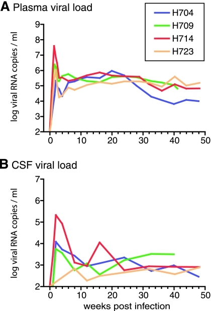
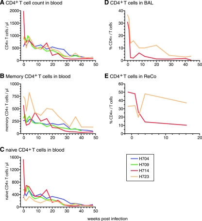
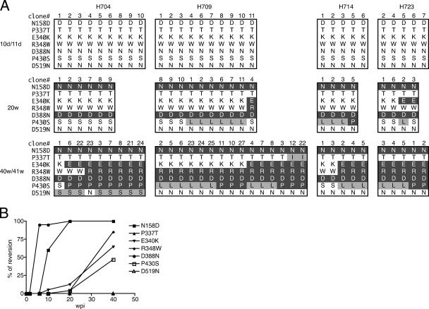
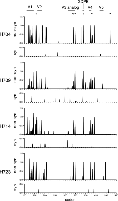
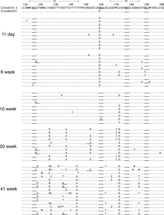
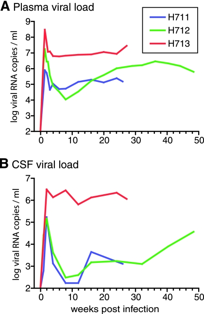
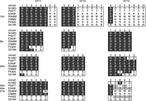
A subset of simian immunodeficiency virus (SIV)-infected macaques progresses rapidly to disease with transient SIV-specific immune responses and high viral loads. Unique SIV variants with convergent Env mutations evolve in these rapid progressor (RP) macaques. To address the pathogenic significance of RP-specific variants, we generated infectious molecular clones from the terminal-phase plasma of an RP macaque. Inoculation of macaques with a representative clone, SIVsmH635FC, resulted in a persistent viremia, comparable to that produced by pathogenic SIVsmE543-3, and a chronic disease with progressive loss of CD4(+) T cells. However, SIVsmH635FC did not reproduce the rapid-disease phenomenon. Molecular analyses of viruses from these macaques revealed rapid reversion to the wild-type SIVsmE543-3 sequence at two RP-specific sites and slower reversion at another three sites. SIVsmH635FC infection was not sufficient to cause rapid progression even following coinoculation with SIVsmE543-3, despite acute depletion of memory CD4(+) T cells. SIVsmH635FC competed efficiently during primary infection in the coinoculated macaques, but SIVsmE543-3 predominated after the development of SIV-specific immune responses. These data suggest that the replication fitness of the RP variant was similar to that of SIVsmE543-3 in a naïve host; however, SIVsmH635FC was at a disadvantage following the development of SIV-specific immune responses. Consistent with these findings, neutralization assays revealed that SIVsmH635FC was highly sensitive to neutralization but that the parental SIVsmE543-3 strain was highly resistant. This study suggests that the evolution of RP-specific variants is the result of replication in a severely immunocompromised host, rather than the direct cause of rapid progression.


Figures

FIG. 1.
FIG. 1.
Viral RNA loads in the plasma (A) and CSF (B) of four SIVsmH635FC-inoculated rhesus macaques (H704, H709, H714, and H723). CSF viral loads of H723 at 1 and 2 wpi were not determined because CSF samples were not available.
FIG. 2.
FIG. 2.
CD4+ T-cell depletion in four SIVsmH635FC-inoculated macaques. The progressive declines in numbers of CD4+ T cells (A), memory (CD95highCD28high or CD95highCD28low) CD4 T cells (B), and naive (CD95lowCD28high) CD4 T cells (C) in blood are shown by absolute numbers. The depletion of CD4+ T cells in mucosal tissues is shown by percentages of CD4+ cells in total T cells from BAL (D) and ReCo (E) samples. BAL and ReCo samples were collected only from H714 and H723.
FIG. 3.
FIG. 3.
Reversion to the parental SIVsmE543-3 sequence in SIVsmH635FC-infected macaques. (A) The env V1-V5 region was amplified by RT-PCR from plasma samples from day 10 or 11 (10d/11d), week 20 (20w), and week 40 or 41 (40w/41w). RT-PCR products were cloned into plasmids and sequenced. Reversion to the SIVsmE543-3 sequence at the seven locations which differ between SIVsmE543-3 and SIVsmH635FC in the env V1-V5 region is shown by amino acids. The seven locations of the amino acid substitutions, namely, N158D, P337T, E340K, R348W, D388N, P430S, and D519N, are shown on the left side. Amino acid residues at these locations of the obtained clones are boxed to represent the sampling date and macaques. Clone numbers are shown at the tops of the boxes. Amino acids that reverted to SIVsmE543-3 amino acids are shown by white letters in a black background. Amino acid changes other than the changes that revert to the SIVsmE543-3 sequence are shaded in gray. (B) The kinetics of reversion to the SIVsmE543-3 sequence are shown. The percentage of reversion was calculated using all the clones from the four SIVsmH635FC-infected macaques at day 10 or 11, week 20, and week 40 or 41 shown in panel A. The percentages of reversion at 6 and 10 weeks were calculated using clones from two macaques, H704 and H709.
FIG. 4.
FIG. 4.
Amino acid alignment of the Env V1-V2 region of clones amplified from plasma samples of the four SIVsmH635FC-infected macaques, H704, H709, H714, and H723, at 40 or 41 wpi. The amino acid sequence of SIVsmE543-3 is shown at the top in the single-letter code, with dots below indicating identical amino acids in aligned sequences. Amino acid substitutions are indicated, gaps are shown by dashes, and PNG sites are underlined.
FIG. 5.
FIG. 5.
Average nonsynonymous and synonymous substitutions of each codon in the env V1-V5 region. Pairwise comparisons with SIVsmH635FC were performed using nucleic acid sequence data of clones amplified from plasma samples of SIVsmH635FC-infected macaques at 40/41 wpi. Numbers of nonsynonymous (non-syn) and synonymous (syn) substitutions per site were calculated with SNAP. The locations of seven nucleotide changes between SIVsmE543-3 and SIVsmH635FC are indicated by inverted triangles at the top. The V1 to V5 regions and GDPE motif are also indicated at the top. The codon numbers of env are shown at the bottom.
FIG. 6.
FIG. 6.
Amino acid alignment of the Env V1-V2 regions of clones amplified from sequential plasma samples of SIVsmH635FC-infected macaque H709. The amino acid sequence of SIVsmE543-3 is shown at the top in the single-letter code, with dots below indicating identical amino acids in aligned sequences. Amino acid substitutions are indicated, gaps are shown by dashes, and PNG sites are shown by underlines.
FIG. 7.
FIG. 7.
Viral RNA loads in the plasma (A) and CSF (B) of three rhesus macaques (H711, H712, and H713) that were coinoculated with SIVsmE543-3 and SIVsmH635FC.
FIG. 8.
FIG. 8.
Kinetics of CD4+ T cells in three rhesus macaques (H711, H712, and H713) that were coinoculated with SIVsmE543-3 and SIVsmH635FC. Decreases in numbers of CD4+ T cells (A), memory (CD95highCD28high or CD95highCD28low) CD4+ T cells (B), and naive (CD95lowCD28high) CD4+ T cells (C) in blood are shown with absolute numbers. The depletion of CD4+ T cells in mucosal tissues is shown by percentages of CD4+ cells in total T cells from BAL (D) and ReCo (E) samples.
FIG. 9.
FIG. 9.
Differentiation of viruses in three macaques coinfected with SIVsmE543-3 and SIVsmH635FC. The env V1-V5 region was amplified by RT-PCR from viral RNA isolated from plasma samples of these macaques at 10 days (10d), 8 weeks (8w), 20 weeks, and 26 weeks (H711), 40 weeks (H712), or 27 weeks (H713) postinoculation. PCR products were cloned into plasmids, and the sequences were analyzed. Amino acids at the seven locations which differ between SIVsmE543-3 and SIVsmH635FC in the Env V1-V5 region are shown as described for Fig. 3A. Amino acids that are identical to those of SIVsmE543-3 are shown by white letters in a black background. Amino acids that are identical to those of SIVsmH635FC are shown by black letters in a white background. Amino acid changes other than changes from SIVsmE543-3 and SIVsmH635FC are shaded.

Similar articles

Cited by

References

    1. Barouch, D. H., J. Kunstman, M. J. Kuroda, J. E. Schmitz, S. Santra, F. W. Peyerl, G. R. Krivulka, K. Beaudry, M. A. Lifton, D. A. Gorgone, D. C. Montefiori, M. G. Lewis, S. M. Wolinsky, and N. L. Letvin. 2002. Eventual AIDS vaccine failure in a rhesus monkey by viral escape from cytotoxic T lymphocytes. Nature 415:335-339. - PubMed
    1. Brown, C. R., M. Czapiga, J. Kabat, Q. Dang, I. Ourmanov, Y. Nishimura, M. A. Martin, and V. M. Hirsch. 2007. Unique pathology in simian immunodeficiency virus-infected rapid-progressor macaques is consistent with a pathogenesis distinct from that of classical AIDS. J. Virol. 81:5594-5606. - PMC - PubMed
    1. Burns, D. P., C. Collignon, and R. C. Desrosiers. 1993. Simian immunodeficiency virus mutants resistant to serum neutralization arise during persistent infection of rhesus monkeys. J. Virol. 67:4104-4113. - PMC - PubMed
    1. Carrington, M., G. W. Nelson, M. P. Martin, T. Kissner, D. Vlahov, J. J. Goedert, R. Kaslow, S. Buchbinder, K. Hoots, and S. J. O'Brien. 1999. HLA and HIV-1: heterozygote advantage and B*35-Cw*04 disadvantage. Science 283:1748-1752. - PubMed
    1. Cecilia, D., V. N. KewalRamani, J. O'Leary, B. Volsky, P. Nyambi, S. Burda, S. Xu, D. R. Littman, and S. Zolla-Pazner. 1998. Neutralization profiles of primary human immunodeficiency virus type 1 isolates in the context of coreceptor usage. J. Virol. 72:6988-6996. - PMC - PubMed

Publication types

MeSH terms