. 2007 Sep;81(17):9469-80.
doi: 10.1128/JVI.00989-07. Epub 2007 Jun 27.

The RNA binding domain of influenza A virus NS1 protein affects secretion of tumor necrosis factor alpha, interleukin-6, and interferon in primary murine tracheal epithelial cells

Affiliations

The RNA binding domain of influenza A virus NS1 protein affects secretion of tumor necrosis factor alpha, interleukin-6, and interferon in primary murine tracheal epithelial cells

Celeste M Newby et al. J Virol. 2007 Sep.

Erratum in

  • J Virol. 2007 Nov;81(22):12717

Abstract

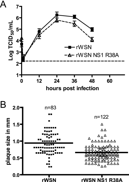
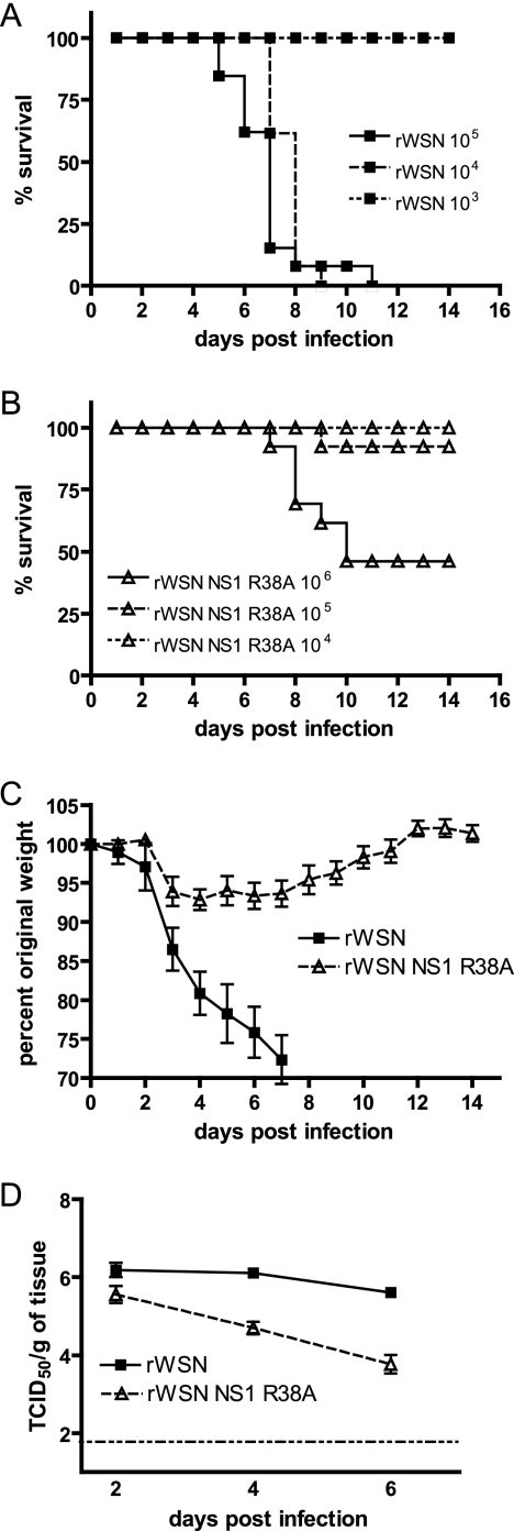
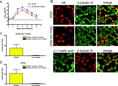
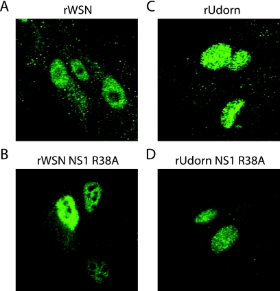
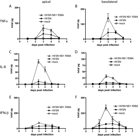
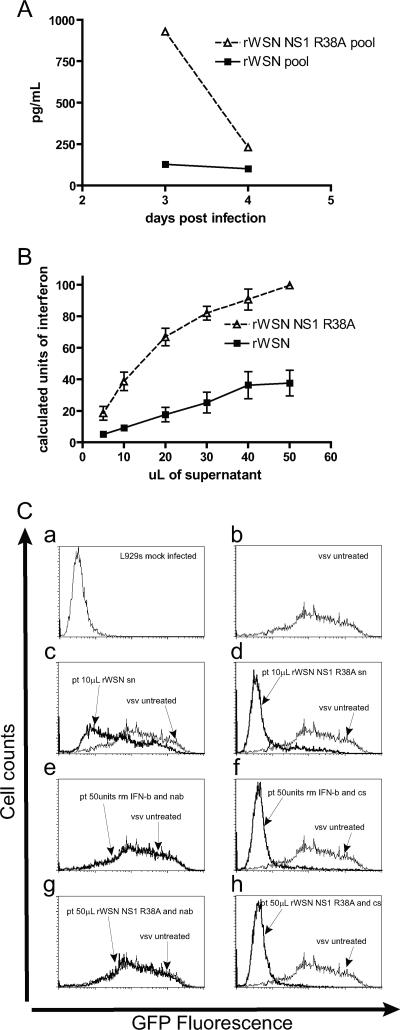
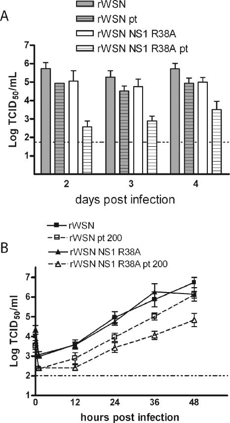
Primary differentiated respiratory epithelial cell cultures closely model the in vivo environment and allow for studies of innate immune responses generated specifically by epithelial cells, the primary cell type infected by human influenza A virus strains. We used primary murine tracheal epithelial cell (mTEC) cultures to investigate antiviral and cytokine responses to influenza A virus infection, focusing on the contribution of the RNA binding domain of the NS1 protein. rWSN NS1 R38A replication is attenuated in mTEC cultures; however, viral antigen is detected predominantly in ciliated cells, similar to wild-type virus. NS1 and NS1 R38A proteins display a primarily cytoplasmic localization in infected mTEC cultures. Increased production of tumor necrosis factor alpha, interleukin-6, and beta interferon is observed during rWSN NS1 R38A infection, and cytokines are secreted in a directional manner. Cytokine pretreatment of mTEC cultures and Vero cells suggest that rWSN NS1 R38A is more sensitive to the presence of antiviral/inflammatory cytokines than wild-type virus. Our results demonstrate that the RNA binding domain is a critical regulator of both cytokine production and cytokine sensitivity during influenza A virus infection of primary tracheal epithelial cells.


Figures

FIG. 1.
FIG. 1.
rWSN NS1 R38A replication in MDCK cells. (A) Multistep growth curve of rWSN and rWSN NS1 R38A. MDCK cells were infected at an MOI of 0.01, supernatants were collected at the times indicated, and infectious virus titers were determined by 50% tissue culture infective dose (TCID50) assays. The data points are average titers from three separate experiments, and error bars indicate standard errors of the means. The dashed horizontal line indicates the limit of detection. (B) Plaque diameters for rWSN and rWSN NS1 R38A on MDCK cells. The solid line indicates the average plaque size for each group.
FIG. 2.
FIG. 2.
rWSN NS1 R38A infection of BALB/c mice. Dose-dependent lethality of (A) rWSN and (B) rWSN NS1 R38A in female BALB/c mice. Virus was administered intranasally with the indicated PFU in a volume of 30 μl, and mice were monitored for mortality for 14 days. (C) Weight loss of rWSN- and rWSN NS1 R38A-infected mice after infection with 1 × 10⁴ PFU. Individual mice were weighed daily, and values are expressed as percents of weight at the time of infection. (D) Lung titers of rWSN- and rWSN NS1 R38A-infected mice after infection with 1 × 10⁴ PFU. Error bars indicate standard errors of the means. The dashed horizontal line indicates the limit of detection. Results were reproduced twice.
FIG. 3.
FIG. 3.
rWSN NS1 R38A replication, antigen expression, and tropism in mTEC cultures. (A) Replication of rWSN and rWSN NS1 R38A in mTECs. Cells were infected at days 10 to 14 after the air-liquid interface with a low MOI (3,600 PFU) and were monitored for the presence of virus in apical and basolateral chambers. Shown are the apical chamber virus titers. Virus was not detected in the basolateral supernatants at any time point. Data points are the averages of four separate experiments, and error bars indicate standard errors of the means. (B) rWSN and rWSN NS1 R38A antigen expression (upper panels) at day 3 postinfection. Red, HA; green, β-tubulin IV. (Lower panel) Expression of α2,3-linked sialic acid (MAA lectin, green) and β-tubulin IV (red) in mTEC cultures. All images were taken at ×63 magnification and are reconstructed from serial optical sections. (C and D) Quantitation of antigen-positive cells for rWSN and rWSN NS1 R38A. Cells in 10 to 15 fields were counted in three separate experiments. The data points are average values with standard errors of the means (error bars).
FIG. 4.
FIG. 4.
NS1 subcellular localization in infected mTEC cultures. NS1 subcellular localization in mTEC cultures at day 3 postinfection. Cells were immunostained with anti-NS1 antibody and the nuclear counterstain TO-PRO-3. (Upper panels) NS1 expression in rWSN-infected cells. (Middle panels) NS1 expression in the majority of rWSN NS1 R38A-infected cells. (Lower panels) Filamentous structures present in a minority of cells infected with rWSN NS1 R38A. All images were taken at ×100 magnification with a 2.5× digital zoom. NS1 images in the top two panels are single-slice images from an 8-μM Z stack with 0.6-μM slices. The filamentous NS1 image (lower panel) shown is a flattened reconstitution from a 10-μM Z stack with 0.6-μM slices in order to highlight the filamentous structures.
FIG. 5.
FIG. 5.
NS1 subcellular localization in MDCK cells. MDCK cells were infected at an MOI of 5 with (A) rWSN, (B) rWSN NS1 R38A, (C) rUdorn, or (D) rUdorn NS1 R38A, fixed at 3 h postinfection, immunostained with anti-NS1 antibody, and analyzed by confocal microscopy. All images were taken at ×63 magnification. The R38A mutation was introduced into the NS segment of rUdorn according to established protocols (112).
FIG. 6.
FIG. 6.
Cytokine production in virus-infected mTEC cultures. Apical and basolateral supernatants were analyzed for the presence of TNF-α, IFN-γ, IL-6, IL-10, MCP-1, and IL-12p70 by cytokine bead array and for the presence of IFN-β by ELISA. Shown are results for TNF-α (A and B), IL-6 (C and D), and IFN-β (E and F) cytokine production in the apical (A, C, and E) and basolateral (B, D, and F) chambers. All other cytokines tested were at levels indistinguishable from those of mock-infected cultures (data not shown). The levels of cytokines present in the apical (left column) and basolateral (right column) chambers were normalized to total picograms to account for the differences in apical (100 μl) and basolateral (500 μl) chamber volumes. Data points are the averages from one experiment, and standard errors of the means (error bars) are shown. Results were replicated at least four times.
FIG. 7.
FIG. 7.
Antiviral activity of basolateral supernatants from rWSN- and rWSN NS1 R38A-infected mTEC cultures. (A) IFN-β ELISA of pooled basolateral samples from rWSN- and rWSN NS1 R38A-infected cultures at days 3 and 4 postinfection. (B) Calculated units of type I IFN activity in rWSN- and rWSN NS1 R38A-infected mTEC culture basolateral supernatants from day 3 postinfection. L929 murine fibroblasts were pretreated overnight with the indicated volumes of basolateral supernatant, infected with VSV-GFP at an MOI of 10, and harvested 10 h postinfection for analysis by flow cytometry. The amount of VSV-GFP inhibition observed with basolateral supernatants was compared to that with a mouse recombinant IFN-β standard curve (not shown) to calculate approximate units of type I interferon. (C) Flow cytometry for VSV-GFP gene expression. (Panel a) Untreated L929 cells. (Panel b) VSV-GFP gene expression on L929 cells. (Panels c and d) Pretreatment with 10 μl of basolateral supernatant from (c) rWSN- or (d) rWSN NS1 R38A-infected mTEC cultures. (Panel e) Pretreatment with 50 U of mouse recombinant IFN-β and type I interferon neutralizing sera. (Panel f) Pretreatment with 50 U of recombinant mouse IFN-β and control sera. (Panel g) Pretreatment with 50 μl of rWSN NS1 R38A basolateral supernatants and type I interferon neutralizing sera. (Panel h) Pretreatment with 50 μl of rWSN NS1 R38A basolateral supernatants and control sera. These experiments were replicated in triplicate using two different pools of basolateral supernatant. pt, pretreatment; sn, supernatant; nab, type I interferon neutralizing antibody; cs, control serum.
FIG. 8.
FIG. 8.
Influenza A virus sensitivity to cytokine pretreatment. (A) Cytokine pretreatment in mTEC cultures. Naïve mTEC cultures were pretreated overnight with 500 μl of the day 3 basolateral supernatants from rWSN NS1 R38A infection. Cells were then infected with rWSN or rWSN NS1 R38A at an MOI of 0.01 (3,600 PFU), and infectious virus production was monitored for 4 days. Infectious virus titers were determined by 50% tissue culture infective dose (TCID50) assay. Shown are the apical titers for each virus. Virus was not detected in the basolateral supernatants during infection or in the supernatants used for pretreatment. Data points are the averages of three replicates, shown with standard errors of the means. Pretreatment had minimal effects on rWSN replication (gray bars versus gray hatched bars), but rWSN NS1 R38A titers were drastically reduced at all times sampled, indicating that the cytokine-rich supernatant was capable of attenuating only rWSN NS1 R38A infection (white bars versus white hatched bars). (B) Vero cells were pretreated overnight with 200 U of recombinant human IFN-β and then infected with rWSN or rWSN NS1 R38A at an MOI of 0.01. Infectious virus titers were determined by TCID50 assay at the times indicated, and standard errors of the means (error bars) are shown. The data was replicated twice.

Similar articles

Cited by

References

    1. Alonso-Caplen, F. V., M. E. Nemeroff, Y. Qiu, and R. M. Krug. 1992. Nucleocytoplasmic transport: the influenza virus NS1 protein regulates the transport of spliced NS2 mRNA and its precursor NS1 mRNA. Genes Dev. 6:255-267. - PubMed
    1. Anonymous. 2004. Update: influenza-associated deaths reported among children aged <18 years—United States, 2003-04 influenza season. Ann. Pharmacother. 38:367-368. - PubMed
    1. Arndt, U., G. Wennemuth, P. Barth, M. Nain, Y. Al-Abed, A. Meinhardt, D. Gemsa, and M. Bacher. 2002. Release of macrophage migration inhibitory factor and CXCL8/interleukin-8 from lung epithelial cells rendered necrotic by influenza A virus infection. J. Virol. 76:9298-9306. - PMC - PubMed
    1. Bergmann, M., A. Garcia-Sastre, E. Carnero, H. Pehamberger, K. Wolff, P. Palese, and T. Muster. 2000. Influenza virus NS1 protein counteracts PKR-mediated inhibition of replication. J. Virol. 74:6203-6206. - PMC - PubMed
    1. Boritz, E., J. Gerlach, J. E. Johnson, and J. K. Rose. 1999. Replication-competent rhabdoviruses with human immunodeficiency virus type 1 coats and green fluorescent protein: entry by a pH-independent pathway. J. Virol. 73:6937-6945. - PMC - PubMed

Publication types

MeSH terms

LinkOut - more resources