. 2007 Sep;81(17):9109-20.
doi: 10.1128/JVI.00827-07. Epub 2007 Jun 27.

Human cytomegalovirus gene expression is silenced by Daxx-mediated intrinsic immune defense in model latent infections established in vitro

Affiliations

Human cytomegalovirus gene expression is silenced by Daxx-mediated intrinsic immune defense in model latent infections established in vitro

Ryan T Saffert et al. J Virol. 2007 Sep.

Abstract

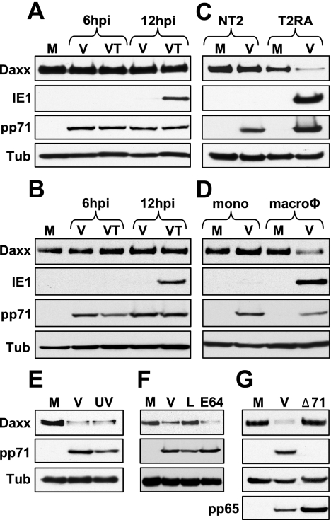
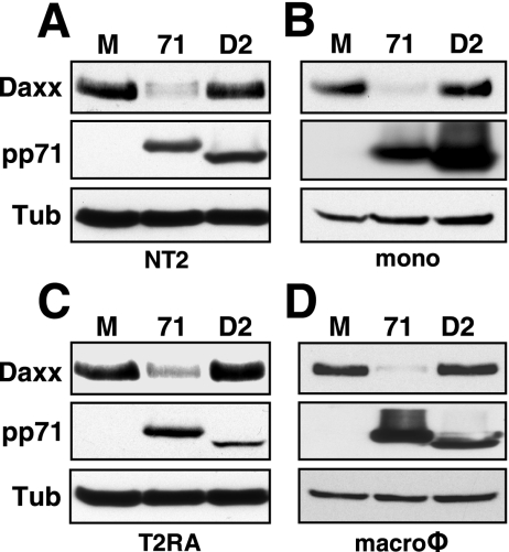
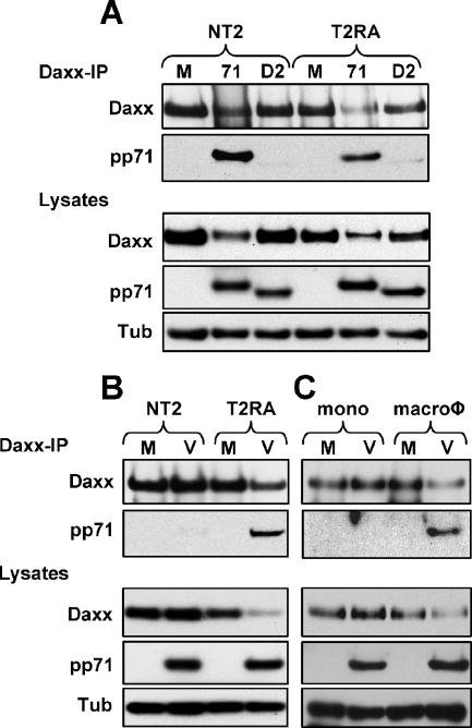
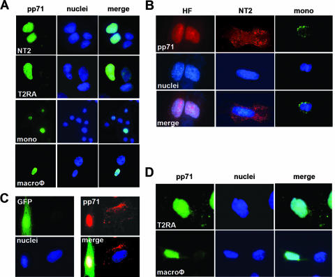
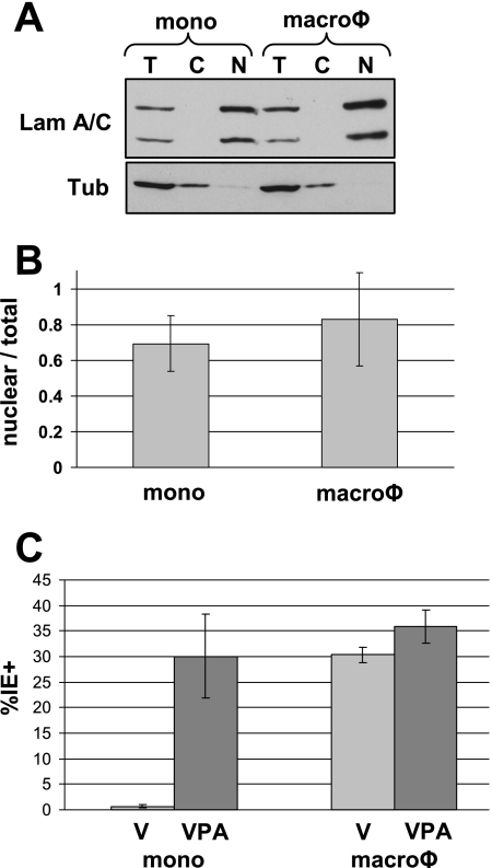
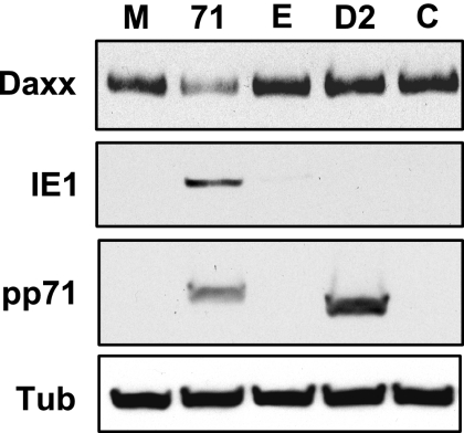
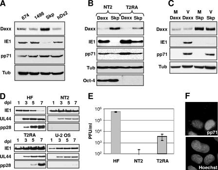
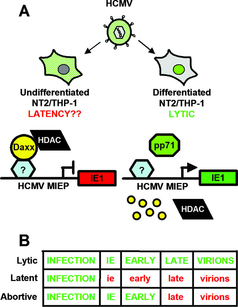
In addition to productive lytic infections, herpesviruses such as human cytomegalovirus (HCMV) establish a reservoir of latently infected cells that permit lifelong colonization of the host. When latency is established, the viral immediate-early (IE) genes that initiate the lytic replication cycle are not expressed. HCMV IE gene expression at the start of a lytic infection is facilitated by the viral pp71 protein, which is delivered to cells by infectious viral particles. pp71 neutralizes the Daxx-mediated cellular intrinsic immune defense that silences IE gene expression by generating a repressive chromatin structure on the viral major IE promoter (MIEP). In naturally latently infected cells and in cells latently infected in vitro, the MIEP also adopts a similar silenced chromatin structure. Here we analyze the role of Daxx in quiescent HCMV infections in vitro that mimic some, but not all, of the characteristics of natural latency. We show that in these "latent-like" infections, the Daxx-mediated defense that represses viral gene expression is not disabled because pp71 and Daxx localize to different cellular compartments. We demonstrate that Daxx is required to establish quiescent HCMV infections in vitro because in cells that would normally foster the establishment of these latent-like infections, the loss of Daxx causes the lytic replication cycle to be initiated. Importantly, the lytic cycle is inefficiently completed, which results in an abortive infection. Our work demonstrates that, in certain cell types, HCMV must silence its own gene expression to establish quiescence and prevent abortive infection and that the virus usurps a Daxx-mediated cellular intrinsic immune defense mechanism to do so. This identifies Daxx as one of the likely multiple viral and cellular determinants in the pathway of HCMV quiescence in vitro, and perhaps in natural latent infections as well.


Figures

FIG. 1.
FIG. 1.
Daxx degradation correlates with IE gene expression. NT2 cells (A) and THP-1 cells (B) were mock infected (M) or infected with HCMV (V) in the absence or presence of trichostatin A (T) at an MOI of 3. Lysates were harvested at the indicated times (hours postinfection [hpi]) and analyzed by Western blotting. Tubulin (Tub) was used as an internal loading control. (C) NT2 and T2RA cells were mock infected (M) or infected with HCMV (V) at an MOI of 3 or 1, respectively. Lysates were harvested at 12 hpi and analyzed by Western blotting. (D) THP-1 monocytes (mono) or THP-1-derived macrophages (macroφ) were mock infected (M) or infected with HCMV (V) at an MOI of 3. Lysates were harvested at 12 hpi and analyzed by Western blotting. (E) T2RA cells were mock infected (M) or infected with HCMV (V) or UV-inactivated HCMV (UV) at an MOI of 3. Lysates were harvested at 12 hpi and analyzed by Western blotting. UV inactivation was confirmed by lack of IE1 gene expression (data not shown). (F) T2RA cells were mock infected (M) or infected with HCMV (V) in the presence of lactacystin (L) or E64 at an MOI of 3. Lysates were harvested at 12 hpi and analyzed by Western blotting. (G) T2RA cells were mock infected (M) or infected with HCMV (V) or the HCMV mutant lacking pp71 (Δ71) at an MOI of either 1 or 0.1, respectively. Lysates were harvested at 12 hpi and analyzed by Western blotting. The tegument protein pp65 served as a marker for viral entry.
FIG. 2.
FIG. 2.
Newly expressed pp71 induces Daxx degradation in undifferentiated and differentiated cells. (A) NT2 cells were mock transduced (lane M) or transduced with an rAD that expresses wild-type pp71 (lane 71) or a mutant pp71 protein (lane D2) which fails to bind Daxx at 2,000 or 4,000 particles per cell (ppc), respectively. Lysates were collected at 24 h posttransduction (hpt) and analyzed by Western blotting. (B) THP-1 monocytes (mono) were mock transduced (lane M) or transduced with the rADs that express pp71 (lane 71) or the pp71 Daxx-binding mutant (lane D2) at 30,000 ppc. Lysates were harvested at 24 hpt and analyzed by Western blotting. (C) Differentiated T2RA cells were mock transduced (lane M) or transduced with an rAD that expresses pp71 (lane 71) or the pp71 Daxx-binding mutant (lane D2) at 300 or 6,000 ppc, respectively. Lysates were harvested at 18 hpt and analyzed by Western blotting. (D) THP-1-derived macrophages (macroφ) were mock transduced (lane M) or transduced with an rAD expressing pp71 (lane 71) or the pp71 Daxx-binding mutant (lane D2) at 5,000 or 30,000 ppc, respectively. Lysates were harvested at 24 hpt and analyzed by Western blotting. Tub, tubulin.
FIG. 3.
FIG. 3.
Tegument-delivered pp71 fails to bind Daxx in undifferentiated NT2 and THP-1 cells. (A) NT2 cells were mock transduced (lane M) or transduced with an rAD expressing either pp71 (lane 71) or the pp71 Daxx-binding mutant (lane D2) at 2,000 or 6,000 particles per cell (ppc), respectively, for 24 h, while differentiated T2RA cells were transduced with pp71 (lane 71) or the pp71 Daxx-binding mutant (lane D2) at 500 or 1,000 ppc, respectively, for 12 h. Lysates were harvested and used in a co-IP/Western blot assay. Lysates were immunoprecipitated with an antibody to Daxx. (B) NT2 or differentiated T2RA cells were mock infected (lane M) or infected with HCMV (lane V) at an MOI of 3 or 1, respectively. Lysates were harvested at 6 hpi and used in a co-IP/Western blot assay. Lysates were immunoprecipitated with an antibody to Daxx. (C) THP-1 monocytes (mono) or THP-1-derived macrophages (macroφ) were mock infected (lane M) or infected with HCMV (lane V) at an MOI of 3 for 12 h. Lysates were harvested and used in a co-IP/Western blot assay. Lysates were immunoprecipitated with an antibody to Daxx. Result from one experiment is shown. Tub, tubulin.
FIG. 4.
FIG. 4.
Recombinant adenovirus-expressed pp71, but not HCMV tegument-delivered pp71, localizes to the nuclei of both undifferentiated and differentiated NT2 and THP-1 cells. (A) NT2, differentiated T2RA cells, THP-1 monocytes (mono), and THP-1-derived macrophages (macroφ) were transduced with the rAD expressing pp71 at 2,000, 1,000, 10,000, or 5,000 particles per cell, respectively. pp71 subcellular localization was visualized by indirect immunofluorescence as described in Materials and Methods. Nuclei were counterstained with Hoechst. pp71 is green, and cellular DNA is blue. (B) HFs, NT2 cells, and THP-1 monocytes (mono) were infected with HCMV at an MOI of 3 for 6 h. Cells on coverslips were stained with the appropriate antibody. Nuclei were counterstained with Hoechst. pp71 is red in HFs and NT2 cells and green in THP-1 monocytes. In all of the panels, cellular DNA is blue. (C) HFs transduced with a retrovirus that expresses GFP were cocultured with NT2 cells grown on coverslips and then infected with HCMV at an MOI of 1. Coverslips were harvested at 10 hpi and assayed by indirect immunofluorescence. GFP is green, pp71 is red, and cellular DNA is blue. (D) Differentiated T2RA cells and THP-1-derived macrophages (macroφ) grown on coverslips were infected with HCMV at an MOI of 1 or 3, respectively. Coverslips were harvested at 8 hpi and assayed by indirect immunofluorescence. pp71 is green, and cellular DNA is blue.
FIG. 5.
FIG. 5.
Viral genomes localize to the nucleus as efficiently in undifferentiated THP-1 cells as in their derivatives. (A) THP-1 monocytes (mono) and macrophages (macroφ) were infected with HCMV at an MOI of 3 for 12 h. Total cell extracts (T) or cytoplasmic (C) and nuclear (N) fractions were analyzed by Western blotting. Lamin (Lam) A/C represents nuclear proteins, while tubulin (Tub) represents cytoplasmic proteins. (B) DNA was harvested from total cell extracts or from isolated nuclei of HCMV-infected THP-1 monocytes (mono) or macrophages (macroφ) and analyzed by real-time Q-PCR. The number of viral genomes detected in the nuclear fraction was divided by the number of viral genomes detected in the total cell extracts. The data represent the average from three independent experiments. (C) THP-1 monocytes (mono) or macrophages (macroφ) were infected with HCMV at an MOI of 3 for 24 h in the absence (V) or presence of VPA. At least 300 nuclei per sample from randomly selected fields were counted, and the data represent an average of the percentage of IE1-positive cells from three independent experiments.
FIG. 6.
FIG. 6.
Preexpression of nuclear pp71 rescues IE1 gene expression. THP-1 monocytes were mock transduced (lane M) or transduced with rAD-EF1α pp71 (lane 71), rAD-EF1α without a transgene (lane E), rAD-CMV pp71 Did2-3 (lane D2), or rAD-CMV without a transgene (lane C) at 2,000 particles per cell for 18 h. Subsequently, all of the cells were infected with HCMV at an MOI of 3 for 24 h. Lysates were harvested and analyzed by Western blotting. Tub, tubulin.
FIG. 7.
FIG. 7.
IE1 is expressed after HCMV infection of undifferentiated cells with reduced Daxx levels, but the infection is abortive. (A) THP-1 monocytes were transfected with siRNA directed at Daxx (674, 1498, or hDx2) or Skp-1 (Skp) for 48 h and then infected with HCMV at an MOI of 3. Lysates were harvested at 24 hpi and analyzed by Western blotting. (B) NT2 or differentiated T2RA cells expressing either shRNA to Daxx or Skp-1 were infected with HCMV at an MOI of 0.5. Lysates were harvested at 12 hpi and analyzed by Western blotting. Oct-4 was used as a marker for the differentiation status of the NT2 cells. (C) THP-1 monocytes expressing either shRNA to Daxx or Skp-1 were mock infected (M) or infected with HCMV (V) at an MOI of 3. Lysates were harvested at 12 hpi and analyzed by Western blotting. (D) HFs, NT2 cells expressing shRNA to Daxx, differentiated T2RA cells expressing shRNA to Daxx, and U-2 OS cells were infected with HCMV at an MOI of 3. Lysates were harvested on the indicated days postinfection (dpi) and analyzed by Western blotting. UL44 served as a marker for early gene expression, and pp28 served as a marker for late gene expression. (E) Supernatants collected at 5 dpi from panel D were used in a plaque assay to determine viral titers. Data collected from two independent experiments are shown with standard deviations. (F) U-2 OS cells were plated on coverslips and infected with HCMV at an MOI of 3. Coverslips were harvested at 8 hpi, and pp71 subcellular localization was visualized by indirect immunofluorescence staining. Nuclei were counterstained with Hoechst. Tub, tubulin.
FIG. 8.
FIG. 8.
Model of differentiation-dependent regulation of pp71 localization, Daxx degradation, and IE gene expression. (A) In undifferentiated NT2/THP-1 monocytes, tegument-delivered pp71 (green) is sequestered in the cytoplasm upon HCMV infection, which allows Daxx to repress IE gene expression through the action of an HDAC. This may mimic what happens when HCMV establishes latency in vivo. However, prior differentiation allows pp71 to localize to the nucleus, induce Daxx degradation, activate viral IE gene expression, and initiate a lytic infection. The small yellow circles represent Daxx degradation. (B) Schematic of the regulation of viral gene expression during lytic, latent, and abortive infections (e.g., when Daxx is absent from cells that would normally establish a latent infection). Green capital letters indicate completion of the particular stage, while red lowercase text indicates failure to complete the phase.

Similar articles

Cited by

References

    1. Albrecht, T., M. Nachtigal, S. C. St. Jeor, and F. Rapp. 1976. Induction of cellular DNA synthesis and increased mitotic activity in Syrian hamster embryo cells abortively infected with human cytomegalovirus. J. Gen. Virol. 30:167-177. - PubMed
    1. Andrews, P. W., I. Damjanov, D. Simon, G. S. Banting, C. Carlin, N. C. Dracopoli, and J. Fogh. 1984. Pluripotent embryonal carcinoma clones derived from the human teratocarcinoma cell line Tera-2. Differentiation in vivo and in vitro. Lab. Investig. 50:147-162. - PubMed
    1. Becker, K. A., L. Florin, C. Sapp, and M. Sapp. 2003. Dissection of human papillomavirus type 33 L2 domains involved in nuclear domains (ND) 10 homing and reorganization. Virology 314:161-167. - PubMed
    1. Bego, M., J. Maciejewski, S. Khaiboullina, G. Pari, and S. St. Jeor. 2005. Characterization of an antisense transcript spanning the UL81-82 locus of human cytomegalovirus. J. Virol. 79:11022-11034. - PMC - PubMed
    1. Bego, M. G., and S. St. Jeor. 2006. Human cytomegalovirus infection of cells of hematopoietic origin: HCMV-induced immunosuppression, immune evasion, and latency. Exp. Hematol. 34:555-570. - PubMed

Publication types

MeSH terms