. 2007 Sep;81(17):9443-50.
doi: 10.1128/JVI.00102-07. Epub 2007 Jun 27.

Respiratory syncytial virus infects and abortively replicates in the lungs in spite of preexisting immunity

Affiliations

Respiratory syncytial virus infects and abortively replicates in the lungs in spite of preexisting immunity

Marina S Boukhvalova et al. J Virol. 2007 Sep.

Abstract

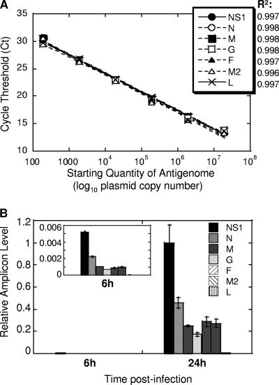
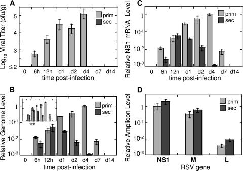
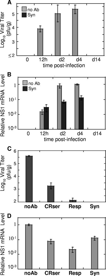
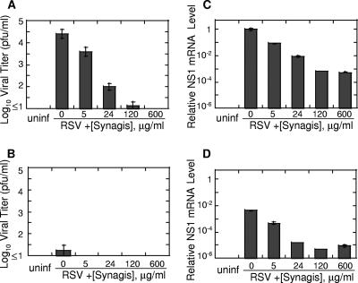
Respiratory syncytial virus (RSV) is a major cause of bronchiolitis and viral pneumonia in young children and a serious health risk in immunocompromised individuals and the elderly. Immunity to RSV is not completely understood. In this work, we established a method for monitoring RSV infection by real-time PCR and applied this method for analysis of RSV replication in vivo in the cotton rat model in naïve animals and in animals rendered immune to RSV by prior RSV infection. We found that even though no virus could be isolated from the lungs of RSV-challenged immune animals, RSV infection in fact took place and an accumulation of viral RNA transcripts was observed. This type of replication, therefore, can be termed "abortive," as RSV is capable of entering the cells in the lungs of immune animals, yet the production of progeny viruses is impaired. Similar patterns of RSV gene expression gradient were observed between naïve and reinfected animals, indicating that the skewing of mRNA gradient of viral gene expression, a mechanism documented during latent infection by other viruses, is not likely to be responsible for abortive replication of RSV during reinfection. We found that passive administration of antibodies to RSV prevents productive infection normally accompanied by viral release in the lung, but it does not prevent abortive replication of the virus. To the best of our knowledge, this is the first evidence of abortive replication of RSV in vivo.


Figures

FIG. 1.
FIG. 1.
Establishing real-time PCR assay of RSV replication. (A) Amplification of various RSV genes from RSV antigenome inserted into a vector. Six 10-fold dilutions of antigenome-containing vector were used to generate a standard curve. CT values were plotted against log10 quantity of antigenome used as a template. Correlation coefficients (R2) of the best fit are shown next to the symbols corresponding to the seven RSV genes analyzed. Each point on this graph includes symbols for all seven genes. (B) Expression of RSV genes in infected A549 cells. A549 cells were infected with RSV at an MOI of 0.1. Six and 24 h after infection, total RNA was extracted from infected cells and reverse transcribed using oligo(dT) primer. Expression of mRNA for each RSV gene indicated was measured by real-time PCR. The relative level of each amplicon was normalized by β-actin mRNA level (also measured by real-time PCR) in the corresponding sample and expressed relative to the level of NS1 amplicon at 24 h. The insert shows gradient of RSV gene expression at 6 h on an adjusted y scale to highlight differences between levels of various amplicons. The results represent the means ± SEM for two different wells per time point.
FIG. 2.
FIG. 2.
RSV replication in vivo: evidence of abortive replication in the reinfection model. CRs were infected with RSV once for primary infection studies or twice (with an interval of 21 days) for secondary infection studies. At the indicated time points after the final challenge, lungs were collected for viral titer determination by plaque assay (A) or for analysis of RSV replication by real-time PCR (B to D). (A) Pulmonary viral titers in CRs with primary RSV infection (prim) and secondary RSV infection (sec). Viral titers were determined by plaque assay as previously described (limit of detection 2 log10 PFU/g) (33). (B) RSV genome and antigenome replication in primary and secondary RSV infection. RNA was extracted from the lungs of infected animals and reversed transcribed using primers complementary to the genomic or antigenomic strand of RSV. Relative genome (and antigenome [insert]) amounts were determined by real-time PCR, normalized by the β-actin mRNA level in the corresponding sample, and expressed relative to the level of genome (or antigenome [insert]) detected in the lungs of animals with primary RSV infection on day 4. (C) Expression of RSV genes during primary and secondary RSV infection. The RNA extracted from animals described above was reverse transcribed using oligo(dT) primer. The expression of NS1 mRNA was measured by real-time PCR, normalized by β-actin as described above, and expressed relative to the level of NS1 mRNA detected in the lungs of animals with primary RSV infection on day 4. (D) Gradient of RSV gene expression at 12 h postinfection in primary and secondary RSV disease. NS1, M, and L amplicon levels were measured by real-time PCR in cDNA prepared using oligo(dT) primer. The expression of each amplicon was normalized by β-actin and expressed relative to the level of NS1 mRNA detected in the lungs of animals with primary RSV infection. The results represent the means ± SEMs for four animals per time point for each primary and secondary infection.
FIG. 3.
FIG. 3.
Abortive replication of RSV in antibody-treated CRs. (A) Naïve CRs were infected with RSV following treatment with anti-RSV antibody palivizumab (Syn) or in the absence of antibody treatment (no Ab). Animals were sacrificed at the indicated times after infection, and viral titers were determined in the lungs by plaque assay. The results represent the means ± SEM for four animals per time point. No virus was detected in the lungs of animals infected with RSV after palivizumab treatment. (B) Real-time PCR analysis of RSV gene expression in the lungs of animals from the experiment described above. Levels of NS1 amplicon were measured by real-time PCR, normalized by the β-actin level in the corresponding organ, and expressed relative to the level of NS1 detected on day 4 in the lungs of naive animals challenged with RSV. (C and D) Abortive RSV replication in the lungs of CRs treated with anti-RSV immune CR serum (CRser) or with RSVIG (Resp). Control groups included CRs infected with RSV in the absence of antibody treatment (noAb) and CRs infected with RSV after palivizumab treatment (Syn). All animals were sacrificed on day 4 postinfection, and lungs were collected for viral titrations (C) and real-time PCR analysis of RSV NS1 expression (D). NS1 levels were expressed relative to the level of NS1 in the “noAb” group. The results represent the means ± SEM for four animals per treatment type.
FIG. 4.
FIG. 4.
Antibodies do not completely prevent infection of epithelial cells and macrophages in vitro. Human A549 alveolar epithelial cells (4 × 10⁵ cells/well) (A and C) and CR peritoneal macrophages (2.5 × 10⁶ cells/well) (B and D) were infected with RSV (MOI of 0.1 to 0.5) preincubated with the indicated amounts of palivizumab (Synagis). Twenty-four hours after infection, cell supernatants were harvested for viral quantification by plaque assay (limit of detection, 1 log10 PFU/g) (A and B), while cells were used for analysis of RSV gene expression (C and D). Total RNA was extracted from infected cells and reverse transcribed using oligo(dT) primer. The expression of mRNA for NS1 gene was measured by real-time PCR, normalized by β-actin mRNA level in the corresponding sample, and expressed relative to the level of NS1 amplicon in A549 cells infected with RSV untreated with antibody (0 μg/ml palivizumab). Uninfected cells (uninf) were used as a negative control. The results represent the means ± SEM for two different wells per treatment type.

Similar articles

Cited by

References

    1. Alexandersen, S., S. Larsen, A. Cohn, A. Uttenthal, R. E. Race, B. Aasted, M. Hansen, and M. E. Bloom. 1989. Passive transfer of antiviral antibodies restricts replication of Aleutian mink disease parvovirus in vivo. J. Virol. 63:9-17. - PMC - PubMed
    1. Arnold, R., B. Konig, H. Galatti, H. Werchau, and W. Konig. 1995. Cytokine (IL-8, IL-6, TNF-α) and soluble TNF receptor-I release from human peripheral blood mononuclear cells after respiratory syncytial virus infection. Immunology 85:364-372. - PMC - PubMed
    1. Bermingham, A., and P. L. Collins. 1999. The M2-2 protein of human respiratory syncytial virus is a regulatory factor involved in the balance between RNA replication and transcription. Proc. Natl. Acad. Sci. USA 96:11259-11264. - PMC - PubMed
    1. Blanco, J. C., L. Pletneva, M. Boukhvalova, J. Y. Richardson, K. A. Harris, and G. A. Prince. 2004. The cotton rat: an underutilized animal model for human infectious diseases can now be exploited using specific reagents to cytokines, chemokines, and interferons. J. Interferon Cytokine Res. 24: 21-28. - PubMed
    1. Brake, D. A. 2003. Parasites and immune responses: memory illusion? DNA Cell Biol. 22:405-419. - PubMed

Publication types

MeSH terms