. 2007 Sep;81(17):9346-56.
doi: 10.1128/JVI.00781-07. Epub 2007 Jun 27.

In vivo and in vitro intragenomic rearrangement of TT viruses

Affiliations

In vivo and in vitro intragenomic rearrangement of TT viruses

Ludmila Leppik et al. J Virol. 2007 Sep.

Abstract

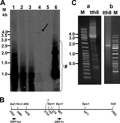
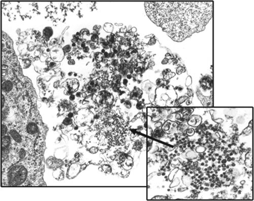
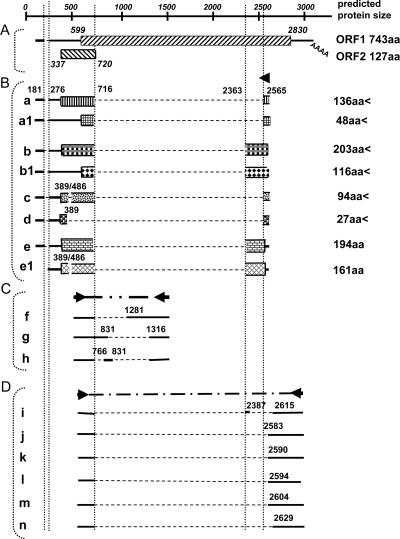
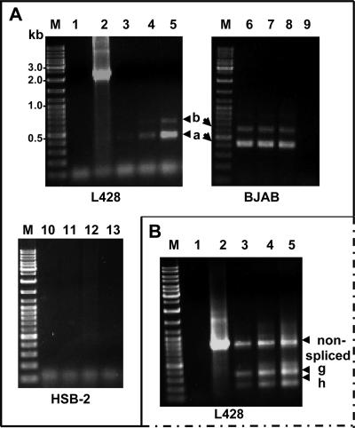
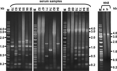
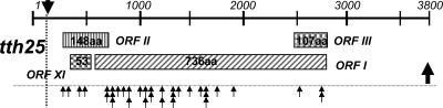
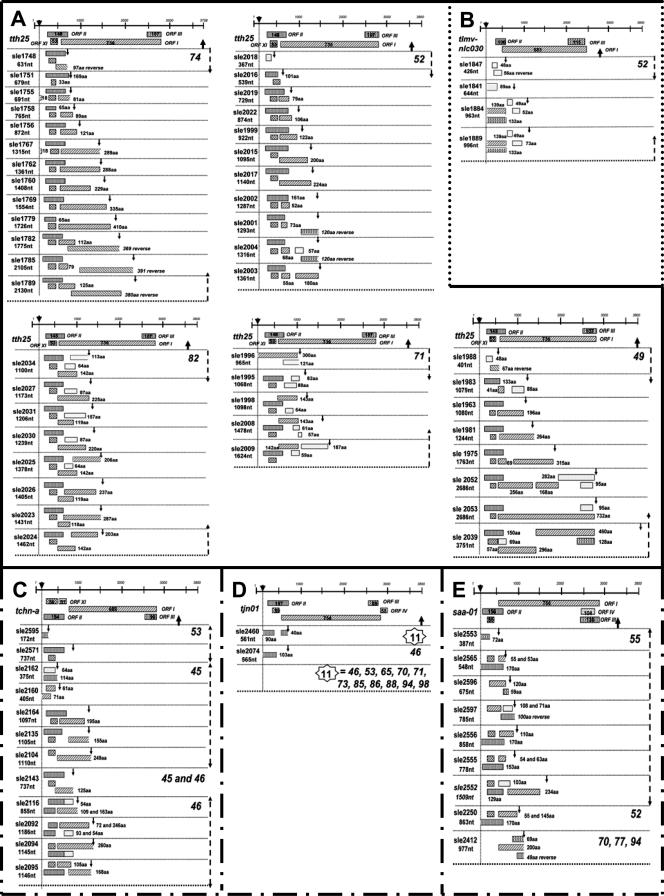
The in vitro replication of the Torque teno virus (TT virus) tth8 full-length genome and particle formation in a Hodgkin's lymphoma-derived cell line after transfection with cloned viral DNA were demonstrated. Analyses of the transcription patterns of tth8 and tth7 TT virus isolates in a number of lymphoma and T-cell leukemia cell lines indicated differential additional splicing events and intragenomic rearrangement generating open reading frames which could not be deducted from the genomic sequence. We also demonstrated the presence of rearranged TT virus genomes in vivo in sera taken from pregnant mothers whose children later developed childhood leukemia, as well as sera from control mothers. Control experiments using religated cloned genomic tth8 DNA mixed with cellular DNA did not result in such subviral molecules. These subviral isolates ranged from 172 bp to full-length TT virus genomes. Possible in vivo selection for specific rearranged molecules was indicated by the presence of one isolate (561 bp) in 11 serum samples. It remains to be clarified whether selected rearranged subviral components resulting from specific TT virus types may contribute to the initiation of disease. These data demonstrate new features of TT viruses suggesting possible similarities to plant viruses of the family Geminiviridae, as well as raise questions about the documented plurality and diversity of anelloviruses.


Figures

FIG. 1.
FIG. 1.
In vitro replication and intragenomic rearrangement of tth8 in the L428 cell line. (A) Southern blot analyses of total cellular DNA harvested at different time points after transfection. DNA was digested with DpnI to fragmentize input viral DNA (#). Hybridization was performed with labeled tth8 DNA. The replication of genomic tth8 DNA (AflII linearized, 3.7 kb; indicated by an arrow) increased from day 2 to day 7 following transfection. Lane 1, 1 day posttransfection; lane 2, 2 days posttransfection; lane 3, 3 days posttransfection; lane 4, 7 days posttransfection; lane 5, negative control with untransfected cells; lane 6, positive control for DpnI digestion of untransfected cellular DNA and religated tth8 genome. M, DNA size marker. (B) Schematic presentation of the tth8 genome after linearization with SalI. The DpnI digestion sites are indicated. ORF1as and ORF1s primers were used to amplify almost-full-length tth8 DNA indicating the recircularization of the linearized tth8 genome used for transfection (data not shown). (C, panel a) PCR amplification of full-length tth8 DNA (3.7 kb), as well as shorter rearranged subviral fragments, 2 days after the transfection of L428 cells. Primers A and B were used for amplification. (C, panel b) Control long-PCR amplification of religated cloned genomic tth8 DNA mixed with L428 cellular DNA. M, DNA size marker.
FIG. 2.
FIG. 2.
Electron micrograph of L428 cells 4 days after transfection with linearized full-length tth8 DNA. TT virus-like particles within a destructed and probably apoptotic cell, measuring approximately 30 to 40 nm in diameter, are visible.
FIG. 3.
FIG. 3.
Schematic presentation of tth7 and tth8 transcripts. Solid lines indicate RNA sequences, and dashed lines represent introns or deleted regions. Nucleotide positions of the junction sites are indicated. Putative ORFs are represented by boxes, and the size of the predicted protein corresponding to each ORF is indicated. Primers used for amplification are shown by arrowheads. aa, amino acids. (A) ORFs as predicted from the full-length transcript of TT virus genomic DNA. (B) mRNA species (a to e1) obtained after 5′-RACE amplification using the 5′NGSP primer. Transcripts a and a1 and transcripts b, b1, and e were identical in length, but sequence analyses resulted in putative proteins of various sizes. (C) mRNA transcripts (f to h) identified by RT-PCR amplification of ORF1 by using the h7g43s and h7g43as primers. (D) Transcripts (i to n) identified by RT-PCR using primers ORF1s and ORF1as.
FIG. 4.
FIG. 4.
Cell lines L428, BJAB, and HSB2 were transfected with tth8 DNA, RNA was isolated 2 days after transfection, and oligo(dT) cDNA synthesis was performed. Regions within ORF1 were amplified. (A) RT-PCR amplification using primers ORF1s and ORF1as. Lanes 1, 9, and 13, negative controls; lane 2, tth8 genomic DNA as a positive control for amplification; lanes 3 to 5, 6 to 8, and 10 to 12, L428, BJAB, and HSB2 samples subjected to 30, 33, and 36 cycles of RT-PCR amplification, respectively. Amplicons indicated the presence of transcripts a and b in L428 and BJAB samples, whereas no transcripts in HSB2 cells were detected. (B) RT-PCR amplification using primers h7g43s and h7g43as (Table 1). Amplicons indicated the presence of a nonspliced transcript, as well as the additional transcripts g and h. Lanes 3, 4, and 5, products from 30, 33, and 36 cycles of RT-PCR amplification, respectively; lane 1, negative control; lane 2, positive control for tth8 genomic DNA amplification; M, DNA size marker.
FIG. 5.
FIG. 5.
Examples of the long-PCR amplification of DNA from individual serum samples using jt34f-1s and jt34f-2as and tth25-1s and tth25-2as. Full-length TT virus genomes (3.5 to 4 kb) as well as off-sized rearranged subviral fragments were amplified. Serum sample numbers are indicated at the top. Long-PCR amplification was controlled by the amplification of religated and rolling-circle-amplified tth8 genomic DNA mixed with placenta DNA (lane a, 102 copies, and lane b, 103 copies of the tth8 genome). M, DNA size marker.
FIG. 6.
FIG. 6.
Breakpoint positions (arrows) in the intragenomic rearranged subviral isolates in relation to the tth25 genome. Boxes indicate the positions of the respective TT virus ORFs.
FIG. 7.
FIG. 7.
Examples of the genomic organization of the rearranged subviral isolates from individual serum samples (numbers are indicated on the right side of each block). The genomic organization of the most closely related known TT virus is indicated at the top of each series. (A) Group most closely related to tth25 (accession no. aj620222); (B) group most closely related to tlmv-nlc030 (accession no. ab0038631); (C) group most closely related to tchn-a (accession no. af345526); (D) group most closely related to tjn01 (accession no. ab028668); and (E) group most closely related to saa-01 (accession no. ab060597). ORFs homologous to known TT virus ORFs are indicated by filled boxes, and those with no identified homology are indicated by unfilled boxes. Deviations from the sizes of known TT virus ORFs are indicated. aa, amino acids.

Similar articles

Cited by

References

    1. Aebi, M., H. Hornig, R. A. Padgett, J. Reiser, and C. Weissmann. 1986. Sequence requirements for splicing of higher eukaryotic nuclear pre-mRNA. Cell 47:555-565. - PubMed
    1. Amin, I., S. Mansoor, L. Amrao, M. Hussain, S. Irum, Y. Zafar, S. E. Bull, and R. W. Briddon. 2006. Mobilization into cotton and spread of a recombinant cotton leaf curl disease satellite. Arch. Virol. 151:2055-2065. - PubMed
    1. Ball, J. K., R. Curran, S. Berridge, A. M. Grabowska, C. L. Jameson, B. J. Thomson, W. L. Irving, and P. M. Sharp. 1999. TT virus sequence heterogeneity in vivo: evidence for co-infection with multiple genetic types. J. Gen. Virol. 80:1759-1768. - PubMed
    1. Biagini, P., P. Gallian, H. Attoui, M. Touinssi, J.-F. Cantaloube, P. de Micco, and X. de Lamballerie. 2001. Genetic analysis of full-length genomes and subgenomic sequences of TT virus-like mini virus human isolates. J. Gen. Virol. 82:379-383. - PubMed
    1. Cacoub, P., E. Rosenthal, V. Gerolami, P. Hausfater, P. Ghillani, Y. Sterkers, V. Thibault, H. Khiri, J. C. Piette, and P. Halfon. 2003. Transfusion-associated TT virus co-infection in patients with hepatitis C virus is associated with type II mixed cryoglobulinemia but not with B-cell non-Hodgkin lymphoma. Clin. Microbiol. Infect. 9:39-44. - PubMed

Publication types

MeSH terms

Associated data

LinkOut - more resources