. 2007 Sep;293(3):R1205-14.
doi: 10.1152/ajpregu.00337.2007. Epub 2007 Jun 27.

PKA phosphorylation of SUR2B subunit underscores vascular KATP channel activation by beta-adrenergic receptors

Affiliations

PKA phosphorylation of SUR2B subunit underscores vascular KATP channel activation by beta-adrenergic receptors

Yun Shi et al. Am J Physiol Regul Integr Comp Physiol. 2007 Sep.

Abstract

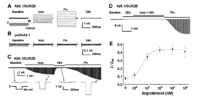
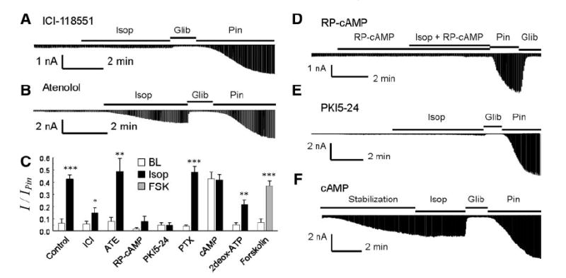
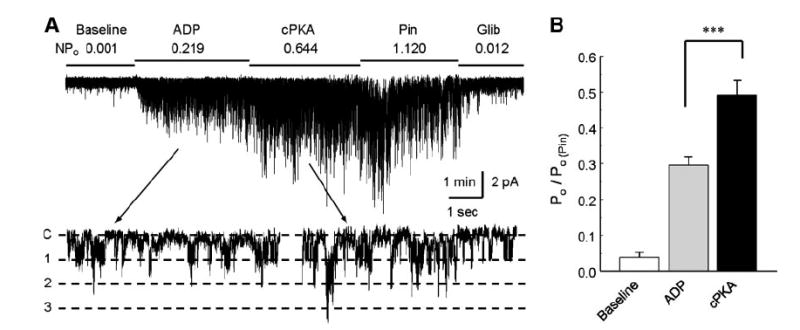
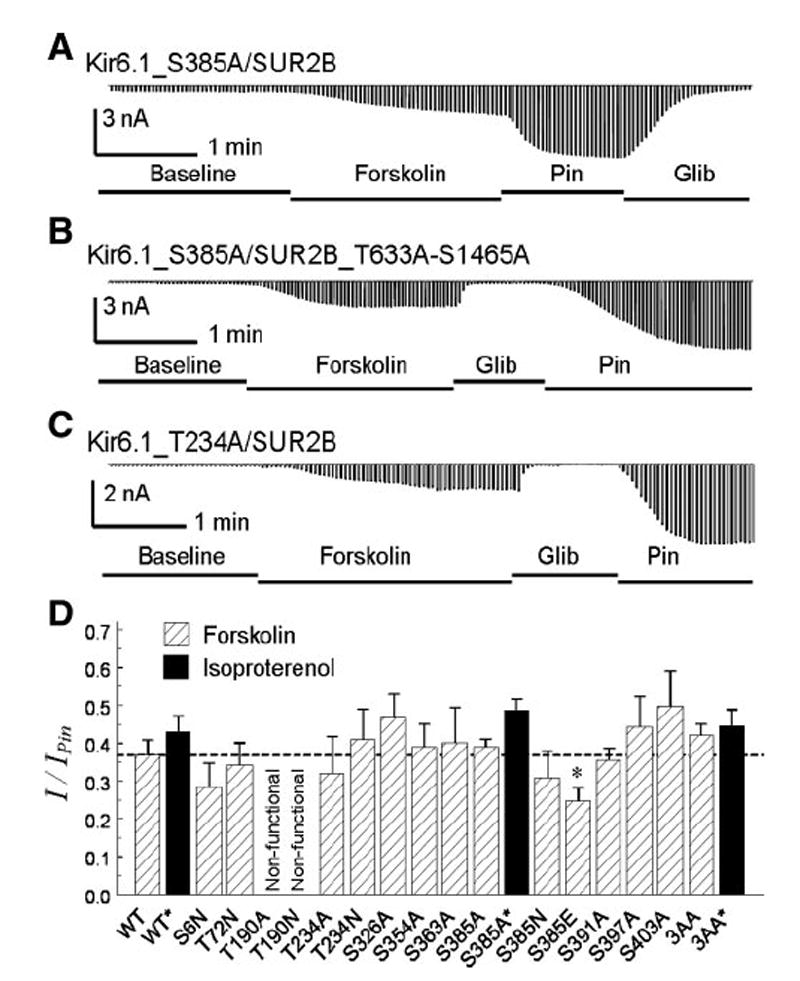
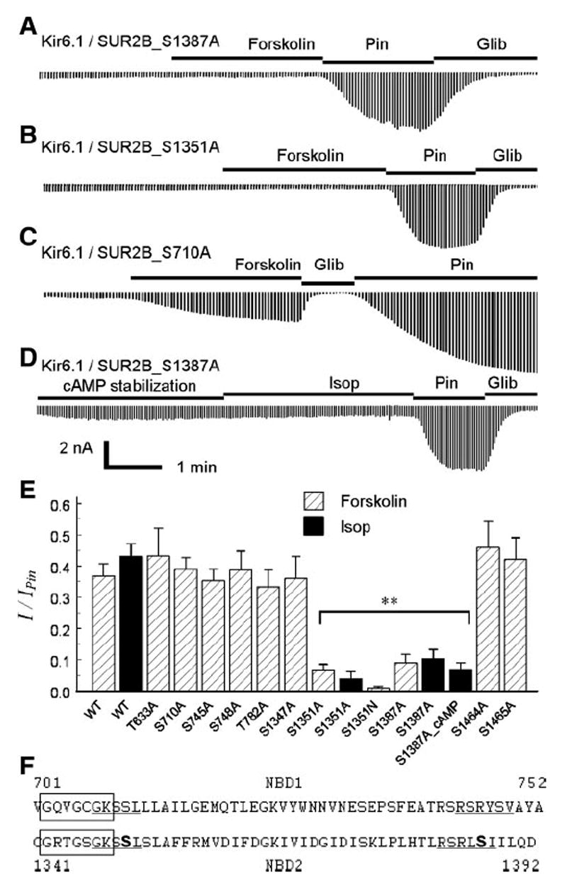
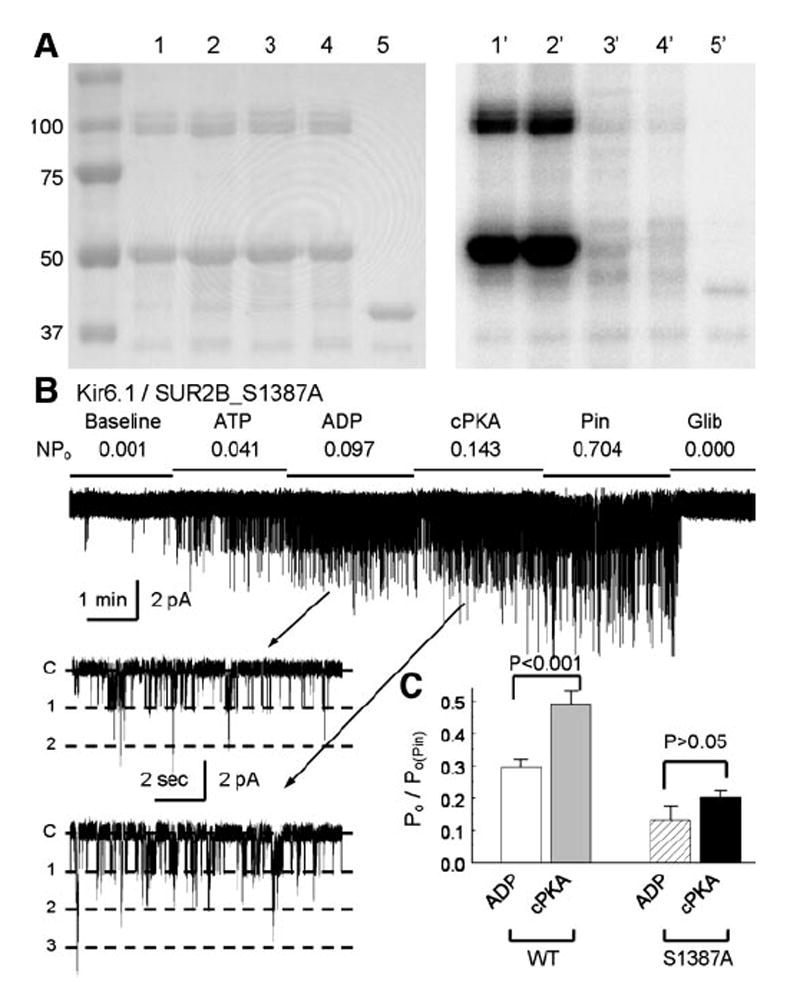
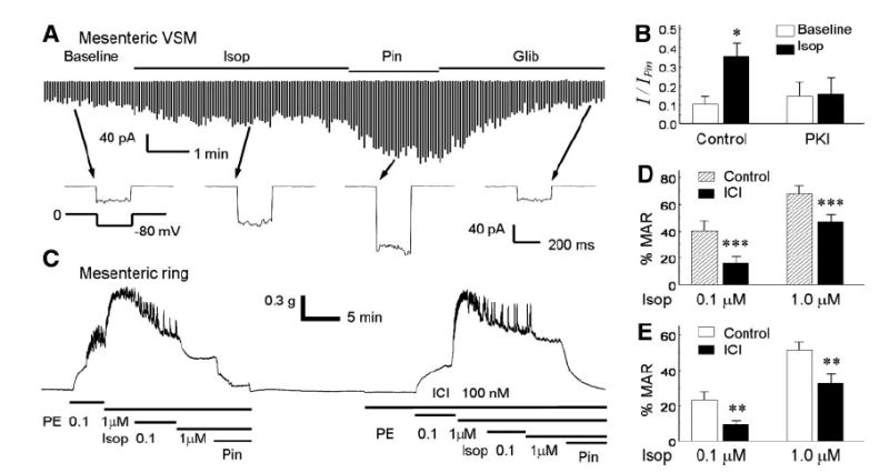
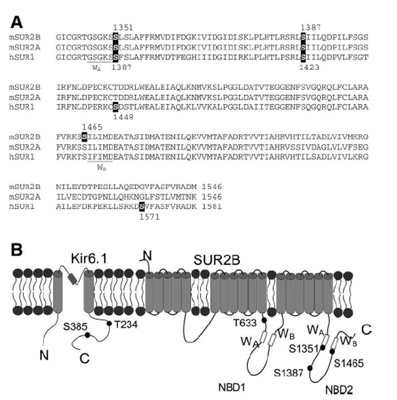
ATP-sensitive K(+) (K(ATP)) channels are activated by several vasodilating hormones and neurotransmitters through the PKA pathway. Here, we show that phosphorylation at Ser1387 of the SUR2B subunit is critical for the channel activation. Experiments were performed in human embryonic kidney (HEK) 293 cells expressing the cloned Kir6.1/SUR2B channel. In whole cell patch, the Kir6.1/SUR2B channel activity was stimulated by isoproterenol via activation of beta(2) receptors. This effect was blocked in the presence of inhibitors for adenylyl cyclase or PKA. Similar channel activation was seen by exposing inside-out patches to the catalytic subunit of PKA. Because none of the previously suggested PKA phosphorylation sites accounted for the channel activation, we performed systematic mutational analysis on Kir6.1 and SUR2B. Two serine residues (Ser1351, Ser1387) located in the NBD2 of SUR2B were critical for the channel activation. In vitro phosphorylation experiments showed that Ser1387 but not Ser1351 was phosphorylated by PKA. The PKA-dependent activation of cell-endogenous K(ATP) channels was observed in acutely dissociated mesenteric smooth myocytes and isolated mesenteric artery rings, where activation of these channels contributed significantly to the isoproterenol-induced vasodilation. Taken together, these results indicate that the Kir6.1/SUR2B channel is a target of beta(2) receptors and that the channel activation relies on PKA phosphorylation of SUR2B at Ser1387.


Figures

Fig. 1
Fig. 1
Kizr6.1/SUR2B channels expressed in human embryonic kidney (HEK)293 cells. A: whole cell currents were recorded from a cell transfected with Kir6.1/SUR2B. Symmetric concentrations of K+ (145 mM) were applied to both sides of cell membranes. The cell was held at 0 mV, and pulse voltages from −120 to 80 mV with a 20-mV increment were applied. The current amplitude increased in response to isoproterenol (Isop; 100 nM). The isoproterenol-activated currents were further activated by pinacidil (Pin; 10 μM) and inhibited by glibenclamide (Glib; 10 μM). B: currents recorded from another cell transfected with the expression vector alone were insensitive to isoproterenol, pinacidil, and glibenclamide. C: time course for the Kir6.1/SUR2B channel modulation. Whole cell currents were recorded with a holding potential at 0 mV and command pulses of −80 mV in every 3 s. After whole cell configuration was formed, the cell was perfused with extracellular solution for a 4-to 6-min baseline recording. Note that the baseline record was shortened in the figure. The currents were strongly activated by isoproterenol, and the maximum activation was reached during 3–4 min of the exposure. The currents were inhibited by glibenclamide (10 μM) and further activated by pinacidil (10 μM). The lower panel shows individual currents produced by a single command pulse. D: in the presence of glibenclamide, isoproterenol failed to activate the Kir6.1/SUR2B channel. E: concentration-dependent activation of Kir6.1/SUR2B currents by isoproterenol. The effect of isoproterenol was measured and normalized between the maximum channel inhibition by 10 μM glibenclamide and the maximum channel activation by 10 μM pinacidil. Baseline currents with no isoproterenol were 7.4 ± 1.9% (n = 18) of full channel activation by pinacidil. Evident activation of the Kir6.1/SUR2B currents was seen with 1 nM (11.5 ± 3.2%, n = 4), and the maximum activation was reached with 100 nM (42. 6 ± 3. 0%, n = 8). Further increase in isoproterenol concentration had no further activation, 1 μM (43.8 ± 5.7%, n = 6) and 10 μM (39.4 ± 5.7%, n = 7). The concentration-current relationship was described using the Hill equation y = 0.074 + 0.355/(1 + (EC50/[Isop])h), where y is normalized Kir6.1/SUR2B currents, [Isop] is isoproterenol concentration, EC50 (4.3 nM) is the Isop concentration for 50% channel activation, and h (1.4) is the Hill coefficient.
Fig. 2
Fig. 2
Dissection of signal pathway for Kir6.1/SUR2B channel activation by isoproterenol. A: in the presence of β2 receptor antagonist ICI-118551 (100 nM), isoproterenol had very little effect on the Kir6.1/SUR2B currents. B: with β1 antagonist atenolol (1 μM), isoproterenol remained to activate the Kir6.1/SUR2B currents. Note that β-AR antagonists were perfused to cells 5 min before and during the isoproterenol exposure. C: currents were normalized to pinacidil and glibenclamide effects. Open bar, baseline current; black bar, isoproterenol; gray bar, forskolin. 2Deox-ATP, 2′,5′-dideoxyadenosine-3′-triphosphate; PTX, pertusis toxin. *P < 0.5; **P < 0.01; ***P < 0.001 (n = 5 to 14). BL, baseline; FSK, forskolin. D: RP-cAMP, a potent PKA inhibitor, was applied in both pipette solution (200 μM) and perfusion solution (100 μM). The current’s activation by isoproterenol was almost completely blocked. E: similar blockade of the channel activation was observed with a PKA inhibitory peptide (PKI5–24, 10 μM) in the pipette solution. F: effect of cAMP (100 μM) in pipette solution on the Kir6.1/SUR2B currents. After formation of whole cell configuration, the current amplitude gradually increased and became plateaued at ~40% of the maximum activation by pinacidil in ~5 min. Application of isoproterenol did not produce further activation of the Kir6.1/SUR2B currents.
Fig. 3
Fig. 3
Augmentation of Kir6.1/SUR2B channel activation by the catalytic subunit of PKA. A: Kir6.1/SUR2B currents were recorded in an inside-out patch obtained from an HEK cell with a holding potential of −60 mV and equal concentrations of K+ applied to both sides of the patch membranes. The channel activity was low in the baseline. Exposure of the internal patch membrane to 1.0 mM ATP and 0.5 mM ADP led to activation of the channels that showed a unitary conductance ~35 pS (lower trace). The channels were further activated with an application of cPKA (100 U/ml) to the internal solution in the presence of same concentrations of ATP and ADP. B: summary of the experiment. Channel activity (NPo) was normalized to the level of pinacidil [Po/Po(Pin)]. The channel activity was rather low at baseline, increased markedly with ADP/ATP, and was further augmented with addition of cPKA. ***P < 0.001 (n = 9 patches).
Fig. 4
Fig. 4
Mutation on potential PKA sites. A: with a mutation of Ser385 to alanine, the Kir6.1_S385A/SUR2 currents were strongly activated by forskolin. B: channel remained to be activated when three potential PKA sites; that is, Ser385 in the Kir6.1, and Ser1465 and Thr633 in the SUR2B subunit, were all mutated to alanine. C: Thr234 mutation on Kir6.1 did not abolish the channel activation by forskolin. D: summary of mutagenesis analysis of potential PKA sites. All mutations were constructed on Kir6.1 expect 3AA in which Ser385 in Kir6.1, and Ser1465/Thr633 in SUR2B were mutated to alanine. All mutant data were obtained from 4–7 cells except two, the 3AA with forskolin and the S385A with isoproterenol, which were obtained from 10 cells. The dashed line indicates the level of WT channel activation by forskolin. *P < 0.05.
Fig. 5
Fig. 5
Identification of PKA phosphorylation sites in SUR2B. Mutants were coexpressed with WT Kir6.1 in HEK cells. A and B: site-specific mutation of Ser1387 and Ser1351 in SUR2B abolishes the channel activation by 10 μM forskolin. C: similar mutation at Ser710 had no effect on the forskolin sensitivity. D: intracellular dialysis of cAMP (100 μM in pipette solution) showed only modest stimulation of the S1387A currents, in sharp contrast to the WT channel shown in Fig. 2F. E: compared with WT, mutations of Ser1351 and Ser1387 caused a loss of channel activation by forskolin. The Ser1351 and Ser1387 mutants failed to be activated by isoproterenol either (**P < 0. 01, n = 4 to 6). F: alignment of amino acid sequences around Walker A in NBD1(top) and NBD2 (bottom). Boxed are Walker A sequences. Ser1351 and Ser1387 are bold, and the proposed PKA consensus sequence is underlined. Similar sequences are seen in NBD1, while both Ser710 and Ser748 are not functional PKA sites.
Fig. 6
Fig. 6
Characterization of Ser1387 in PKA phosphorylation. A: short peptide in SUR2B (residues 1308–1399) was fused to C-terminus of MBP. Site-specific mutations of S1351A and/or S1387A were created on the peptide. After purification with amylose-affinitive beads, correct peptides were revealed in SDS-PAGE gel (left). A 50-kDa band is seen in lanes 14 containing WT, S1351A, S1387A, and S1351A/S1387A double mutations, respectively. Lane 5, MBP protein. Another band of ~100 kDa is also seen, suggesting dimerization of the fusion peptides. Autoradiograph with 32P-γ-labeled ATP after an 8-h exposure (right) showed positive labeling of the WT and S1351A but not the S1387A, and S1351A/S1387A peptides and the control MBP. B and C: compared with the WT channel, the S1387A mutant was slightly activated by the catalytic subunit of PKA (n = 5). Note that the S1387A currents with cPKA exposure were smaller than the baseline level of the WT channel.
Fig. 7
Fig. 7
Effects of isoproterenol on vascular smooth muscles. A: whole-cell currents were recorded from a vascular smooth myocyte (VSM) acutely dissociated from the mesenteric artery. Exposure to 100 nM isoproterenol activated the currents that were further activated by 10 μM pinacidil. Bottom: current traces recorded with a single voltage protocol. B: isopoterenol-activated currents were not seen in the presence of PKI5–24 (PKI, 10 μM) in the pipette solution. C: in a mesenteric ring, phenylephrine (PE) produced vasoconstriction that was reversed by isoproterenol. Such vasorelaxation effects were markedly attenuated in the presence of a β2-AR antagonist ICI-118551 (ICI). Note that pinacidil can completely relax the mesenteric ring. D: summary of vasorelaxation effects of isoproterenol on the PE-induced vasoconstriction with and without ICI on endothelium-intact rings (n = 7). E: similar vasorelaxation was observed in endothelium-denuded rings (n = 6). **P < 0.01; ***P < 0.001.
Fig. 8
Fig. 8
The sites important for PKA regulation on KATP channels. A: alignment of NBD2 of mouse SUR2A, SUR2B (residue 1339-end), and human SUR1 (residue 1376-end). The nucleotide binding Walker A (WA) and Walker B (WB) are underlined. The S1351 that is found important for PKA activation and PKA phosphorylation site (S1387) in SUR2B, and the conserved sites in SUR2A and SUR1 are highlighted in reverse form. The previously suggested PKA site (S1465) in SUR2B and sites (S1448 and S1571) in SUR1 are also highlighted. B: schematic representation of Kir6.1 and SUR2B. The nucleotide binding domain 1 (NBD1) and 2 (NBD2) and Walker A (WA) and Walker B (WB) and N, C terminus are illustrated. The relative positions of important sites (Kir6.1_Thr234, Ser385 and SUR2B_Thr633, Ser1351, Ser1387, and Ser1465) are marked. Note the Kir6.1_Thr234 and Ser385 are also the corresponding sites of Kir6.2_Thr224 and Ser372).

Similar articles

Cited by

References

    1. Aguilar-Bryan L, Bryan J. Molecular biology of adenosine triphosphate-sensitive potassium channels. Endocr Rev. 1999;20:101–135. - PubMed
    1. Beguin P, Nagashima K, Nishimura M, Gonoi T, Seino S. PKA-mediated phosphorylation of the human KATP channel: separate roles of Kir6.2 and SUR1 subunit phosphorylation. EMBO J. 1999;18:4722–4732. - PMC - PubMed
    1. Blom N, Sicheritz-Ponten T, Gupta R, Gammeltoft S, Brunak S. Prediction of post-translational glycosylation and phosphorylation of proteins from the amino acid sequence. Proteomics. 2004;4:1633–1649. - PubMed
    1. Cao K, Tang GH, Hu DH, Wang R. Molecular basis of ATP-sensitive K+ channels in rat vascular smooth muscles. Biochem Biophys Res Commun. 2002;296:463–469. - PubMed
    1. Chang HY. The involvement of ATP-sensitive potassium channels in beta2-adrenoceptor agonist-induced vasodilatation on rat diaphragmatic microcirculation. Br J Pharmacol. 1997;121:1024–1030. - PMC - PubMed

Publication types

MeSH terms

LinkOut - more resources