

Spectrum of clinically relevant Exophiala species in the United States
- PMID: 17596364
- PMCID: PMC2168524
- DOI: 10.1128/JCM.02012-06
Spectrum of clinically relevant Exophiala species in the United States
Abstract
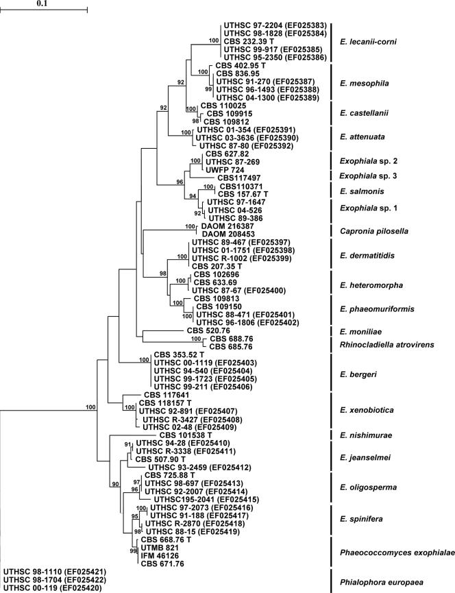
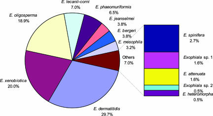
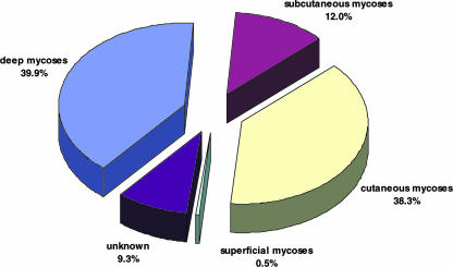
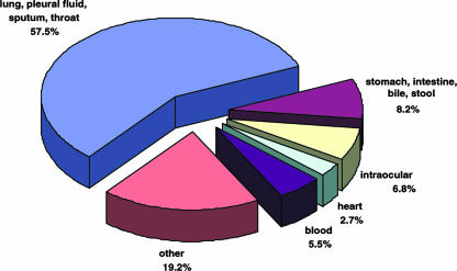
Numerous members of the genus Exophiala are potential agents of human and animal mycoses. The majority of these infections are cutaneous and superficial, but also fatal systemic infections are known. We re-identified 188 clinical isolates from the United States, which had a preliminary morphological identification of Exophiala species, by sequencing internal transcribed spacer (ITS) region of the rRNA. Molecular identifications of the strains were as follows, in order of frequency: 55 E. dermatitidis (29.3%), 37 E. xenobiotica (19.7%), 35 E. oligosperma (18.6%), 13 E. lecanii-corni (6.9%), 12 E. phaeomuriformis (6.4%), 7 E. jeanselmei (3.7%), 7 E. bergeri (3.7%), 6 E. mesophila (3.2%), 5 E. spinifera (2.7%), 3 Exophiala sp. 1 (1.6%), 3 E. attenuata (1.6%), 3 Phialophora europaea (1.3%), 1 E. heteromorpha (0.5%), and 1 Exophiala sp. 2 (0.5%) strains. Exophiala strains were repeatedly isolated from deep infections (39.9%) involving lung, pleural fluid, sputum, digestive organs (stomach, intestines, bile), heart, brain, spleen, bone marrow, blood, dialysis fluid, lymph node, joint, breast, middle ear, throat, and intraocular tissues. About 38.3% of the Exophiala spp. strains were agents of cutaneous infections including skin, mucous membranes, nail, and corneal epithelium lesions. The other strains caused superficial infections (0.5%, including hair) or subcutaneous infection (12.0%, including paranasal sinusitis, mycetoma, and subcutaneous cyst). The systemic infections were preponderantly caused by E. dermatitidis, E. oligosperma, E. phaeomuriformis, E. xenobiotica, and E. lecanii-corni. Strains of E. bergeri, E. spinifera, E. jeanselmei, E. mesophila, and E. attenuata mainly induced cutaneous and subcutaneous infections. Since relatively few unknown ITS motifs were encountered, we suppose that the list of opportunistic Exophiala species in temperate climates is nearing completion, but a number of species still have to be described.
Figures
References
-
- Al-Obaid, I., S. Ahmad, Z. U. Khan, B. Dinesh, and H. M. Hejab. 2006. Catheter-associated fungemia due to Exophiala oligosperma in a leukemic child and review of fungemia cases caused by Exophiala species. Eur. J. Clin. Microbiol. Infect. Dis. 25:729-732. - PubMed
-
- Anonymous. 2002. Exophiala infection from contaminated injectable steroids prepared by a compounding pharmacy-United States, July-November 2002. JAMA 289:291-293. - PubMed
-
- Blaschke-Hellmessen, R. 1996. Fluconazole and itraconazole susceptibility testing with clinical yeast isolates and algae of the genus Prototheca by means of the Etest. Mycoses 39(Suppl. 2):39-43. - PubMed
-
- Campos-Takaki, G. M., and M. L. Jardim. 1994. Report of chronic subcutaneous abscesses caused by Exophiala spinifera. Mycopathologia 127:73-76. - PubMed
Publication types
MeSH terms
Substances
LinkOut - more resources
Full Text Sources
Miscellaneous
