. 2007 Jun 27;27(26):6956-64.
doi: 10.1523/JNEUROSCI.2584-06.2007.

Serotonin activates the hypothalamic-pituitary-adrenal axis via serotonin 2C receptor stimulation

Affiliations

Serotonin activates the hypothalamic-pituitary-adrenal axis via serotonin 2C receptor stimulation

Lora K Heisler et al. J Neurosci. .

Abstract

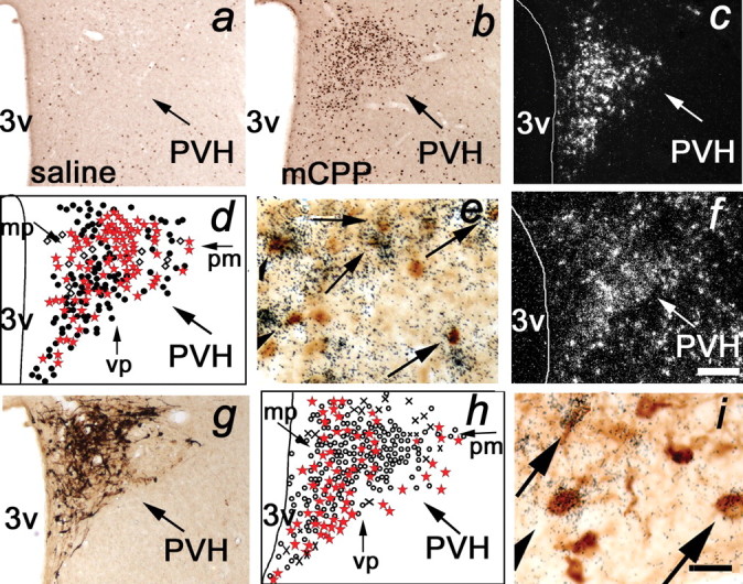
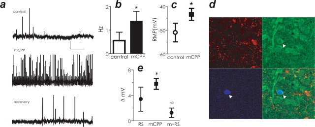
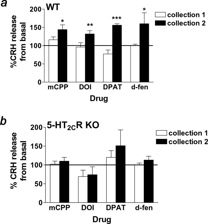
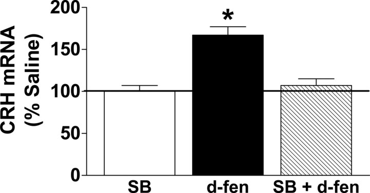
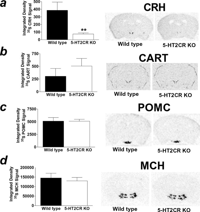
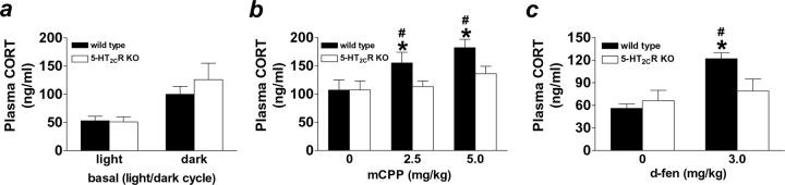
The dynamic interplay between serotonin [5-hydroxytryptamine (5-HT)] neurotransmission and the hypothalamic-pituitary-adrenal (HPA) axis has been extensively studied over the past 30 years, but the underlying mechanism of this interaction has not been defined. A possibility receiving little attention is that 5-HT regulates upstream corticotropin-releasing hormone (CRH) signaling systems via activation of serotonin 2C receptors (5-HT(2C)Rs) in the paraventricular nucleus of the hypothalamus (PVH). Through complementary approaches in wild-type rodents and 5-HT(2C)R-deficient mice, we determined that 5-HT(2C)Rs are necessary for 5-HT-induced HPA axis activation. We used laser-capture PVH microdissection followed by microarray analysis to compare the expression of 13 5-HTRs. Only 5-HT(2C)R and 5-HT(1D)R transcripts were consistently identified as present in the PVH, and of these, the 5-HT(2C)R was expressed at a substantially higher level. The abundant expression of 5-HT(2C)Rs in the PVH was confirmed with in situ hybridization histochemistry. Dual-neurohistochemical labeling revealed that approximately one-half of PVH CRH-containing neurons coexpressed 5-HT(2C)R mRNA. We observed that PVH CRH neurons consistently depolarized in the presence of a high-affinity 5-HT(2C)R agonist, an effect blocked by a 5-HT(2C)R antagonist. Supporting the importance of 5-HT(2C)Rs in CRH neuronal activity, genetic inactivation of 5-HT(2C)Rs produced a downregulation of CRH mRNA and blunted CRH and corticosterone release after 5-HT compound administration. These findings thus provide a mechanistic explanation for the longstanding observation of HPA axis stimulation in response to 5-HT and thereby give insight into the neural circuitry mediating the complex neuroendocrine responses to stress.


Figures

Figure 1.
Figure 1.
mCPP and d-fen activated PVH CRH-containing neurons expressing 5-HT2CRs in vivo. a, b, Compared with saline (a), mCPP (b; 2.5 mg/kg, i.v.) induced a significant increase in FOS-IR (brown nuclear stain) in parvocellular PVH neurons. c–e, mCPP induced FOS-IR in PVH CRH mRNA-containing neurons. c, CRH mRNA (cluster of white grains) is expressed in parvocellular PVH neurons. d, Schematic representation of PVH CRH mRNA and FOS-IR coexpression. Open diamonds, CRH mRNA; filled circles, FOS-IR; red stars, coexpression of CRH mRNA and FOS-IR. e, Arrows identify coexpression of CRH mRNA (cluster of black grains) and FOS-IR (brown nuclear stain). f, 5-HT2CR mRNA (cluster of white grains) is expressed in parvocellular PVH neurons. g–i, 5-HT2CR mRNA is coexpressed with PVH CRH-IR-containing neurons. g, PVH CRH-IR (brown cytoplasm stain) expression. h, Schematic of PVH 5-HT2CR mRNA and CRH-IR coexpression. Circles, CRH-IR; crosses, 5-HT2CR mRNA; red stars, coexpression of CRH-IR and 5-HT2CR mRNA. i, Arrows identify coexpression of 5-HT2CR mRNA (cluster of black grains) and CRH-IR (brown cytoplasm stain). 3v, Third ventricle; mp, medial parvocellular division; pm, posterior magnocellular division; vp, ventral parvocellular division. Scale bars: (in f) a–d, f–h, 200 μm; (in f) e, 20 μm; i, 30 μm.
Figure 2.
Figure 2.
mCPP depolarized and increased the firing rate of CRH-containing neurons in the PVH. a, mCPP depolarized mpPVH neurons. Membrane potential of mpPVH neuron recorded in control saline (top), in the presence of mCPP (middle), and after a 15 min washout (bottom). Calibration bars: 20 mV, 10 s. b, mCPP increased the firing rate of mpPVH CRH-containing neurons compared with control conditions. c, mCPP depolarized mpPVH CRH-containing neurons compared with control conditions. RMP, Resting membrane potential. d, Confirmation that recordings were made from CRH-containing neurons (arrowhead indicates recorded cell). Top left, TRH immunoreactivity; top right, CRH immunoreactivity; bottom left, biocytin histochemistry; bottom right, three images merged. e, 5-HT2CR antagonist RS102221 (RS) did not significantly change membrane potential compared with control. In contrast, mCPP significantly changed membrane potential, and this effect was blocked by treatment with RS. Data are expressed as mean ± SEM. *p < 0.05 compared with control; p < 0.05 compared with mCPP.
Figure 3.
Figure 3.
Genetic 5-HT2CR inactivation abolished 5-HT compound-stimulated CRH release. a, b, mCPP (1.0 μm), DOI (10.0 μm), 8-OH-DPAT (1.0 μm), and d-fen (1.0 μm) stimulated the in vitro release of CRH 15–45 min after drug infusion (black bars; collection 2) in wild-type mice (WT; a) but not 5-HT2CR knock-out littermates (5-HT2CR KO; b) relative to the basal collection period in hypothalamic extracts. Zero to fifteen minutes after drug infusion (white bars; collection 1), CRH release was not different from basal release in either genotype. Data are expressed as mean ± SEM. *p < 0.05; **p < 0.01; ***p < 0.001 compared with basal CRH release using paired t test.
Figure 4.
Figure 4.
Pharmacological 5-HT2CR blockade abolished d-fen-induced enhancement of CRH mRNA expression. Acute treatment with the 5-HT2CR antagonist SB242084 (SB; 1.0 mg/kg, i.p.; white bar) produced no effect on CRH mRNA compared with 0.9% saline treatment. Relative to SB treatment, d-fen (3.0 mg/kg, i.p.; black bar) significantly increased the percentage of CRH mRNA expression in the hypothalamus as measured by real-time quantitative PCR, and pretreatment with SB (1.0 mg/kg, i.p.; striped bar) abolished this effect. Data are expressed as mean ± SEM. *p < 0.05, d-fen compared with all other conditions.
Figure 5.
Figure 5.
5-HT2CR knock-out mice displayed downregulated PVH CRH mRNA but similar PVH CART, ARC POMC, and LHA MCH mRNA expression compared with wild-type littermates. In situ hybridization of 35S-labeled CRH, CART, POMC, and MCH antisense probes was performed in adjacent coronal sections of wild-type (black bar) and 5-HT2CR knock-out (KO; white bar) littermate mouse brains. a, 5-HT2CR KO mice displayed reduced PVH CRH mRNA compared with wild-type littermates as determined by integrated density analysis of 35S-labeled CRH. The autoradiogram illustrates representative sections 0.82 caudal to bregma. b–d, In contrast, 5-HT2CR KO and wild-type mice exhibited comparable levels of PVH 35S-labeled CART (b; autoradiogram illustrates representative sections 0.82 caudal to bregma), similar levels of ARC 35S-labeled POMC (c; autoradiogram illustrates representative sections 1.70 caudal to bregma), and comparable levels of LHA 35S-labeled MCH (d; autoradiogram illustrates representative sections 1.94 caudal to bregma). Data are expressed as mean ± SEM. **p < 0.01, wild-type versus 5-HT2CR KO mice.
Figure 6.
Figure 6.
5-HT2CR knock-out mice displayed normal circadian CORT but did not respond to mCPP- or d-fen-stimulated CORT release. a, Wild-type (black bars) and 5-HT2CR knock-out (KO; white bars) mice displayed comparable levels of CORT measured in the plasma 1 h after the onset of the light and dark cycles. In contrast, a significant genotypic difference in CORT release was observed after HPA axis stimulation. b, c, Both mCPP (b) and d-fen (c) significantly increased in vivo release of CORT in wild-type mice but were ineffective in 5-HT2CR KO littermates. Data are expressed as mean ± SEM. #p < 0.05, drug versus 0.9% saline treatment; *p < 0.05, wild-type versus 5-HT2CR KO mice.

Similar articles

Cited by

References

    1. Bagdy G, Calogero AE, Aulakh CS, Szemeredi K, Murphy DL. Long-term cortisol treatment impairs behavioral and neuroendocrine responses to 5-HT1 agonists in the rat. Neuroendocrinology. 1989;50:241–247. - PubMed
    1. Bhatnagar S, Sun LM, Raber J, Maren S, Julius D, Dallman MF. Changes in anxiety-related behaviors and hypothalamic-pituitary-adrenal activity in mice lacking the 5-HT-3A receptor. Physiol Behav. 2004;81:545–555. - PubMed
    1. Brady LS, Whitfield HJ, Jr, Fox RJ, Gold PW, Herkenham M. Long-term antidepressant administration alters corticotropin-releasing hormone, tyrosine hydroxylase, and mineralocorticoid receptor gene expression in rat brain. Therapeutic implications. J Clin Invest. 1991;87:831–837. - PMC - PubMed
    1. Broocks A, Bandelow B, George A, Jestrabeck C, Opitz M, Bartmann U, Gleiter CH, Meineke I, Roed IS, Ruther E, Hajak G. Increased psychological responses and divergent neuroendocrine responses to m-CPP and ipsapirone in patients with panic disorder. Int Clin Psychopharmacol. 2000;15:153–161. - PubMed
    1. Calogero AE, Bernardini R, Margioris AN, Bagdy G, Gallucci WT, Munson PJ, Tamarkin L, Tomai TP, Brady L, Gold PW, Chrousos GP. Effects of serotonergic agonists and antagonists on corticotropin-releasing hormone secretion by explanted rat hypothalami. Peptides. 1989;10:189–200. - PubMed

Publication types

MeSH terms